HLA-DRB1 Mouse anti-Human, Clone: LN-3, Abnova™
Manufacturer: Abnova Corporation
Select a Size
| Pack Size | SKU | Availability | Price |
|---|---|---|---|
| Each of 1 | 89-934-275-Each-of-1 | In Stock | ₹ 67,017.00 |
89-934-275 - Each of 1
In Stock
Quantity
1
Base Price: ₹ 67,017.00
GST (18%): ₹ 12,063.06
Total Price: ₹ 79,080.06
Antigen
HLA-DRB1
Classification
Monoclonal
Conjugate
Unconjugated
Dilution
Flow Cytometry (0.5-1 ug/106cells in 0.1 mL) Immunofluorescence (0.5-1 ug/mL) Immunohistochemistry (Formalin/PFA-fixed paraffin-embedded sections) (0.25-0.5 ug/mL) Western Blot (0.5-1 ug/mL) The optimal working dilution should be d
Gene Alias
DRB1/FLJ76359/HLA-DR1B/HLA-DRB/HLA-DRB1*/SS1
Host Species
Mouse
Purification Method
Protein A/G purification
Regulatory Status
RUO
Gene ID (Entrez)
3123
Target Species
Human
Form
Liquid
Applications
Flow Cytometry, Immunofluorescence, Immunohistochemistry (PFA fixed), Western Blot
Clone
LN-3
Description
Mouse monoclonal antibody raised against native human HLA-DRB1.
Formulation
In 10mM PBS.
Gene Symbols
HLA-DRB1
Immunogen
Activated human peripheral blood mononuclear cells.
Quantity
100 μg
Primary or Secondary
Primary
Test Specificity
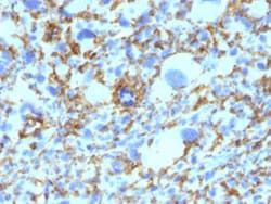
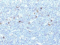
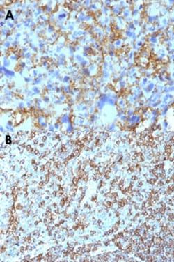
This monoclonal antibody does not cross react with HLA-DP and HLA-DQ. HLA-DR is a heterodimeric cell surface glycoprotein comprised of a 36 kDa alpha (heavy) chain and a 28 kDa beta (light) chain. It is expressed on B cells, activated T cells, monocytes/macrophages, dendritic cells and other non-professional APCs. In conjunction with the CD3/TCR complex and CD4 molecules, HLA-DR is critical for efficient peptide presentation to CD4+T cells. It is an excellent histiocytic marker in paraffin sections producing intense staining. True histiocytic neoplasms are similarly positive. HLA-DR antigens also occur on a variety of epithelial cells and their corresponding neoplastic counterparts. Loss of HLA-DR expression is related to tumor microenvironment and predicts adverse outcome in diffuse large B cell lymphoma.
Content And Storage
Store at -20 to -80°C.Aliquot to avoid repeated freezing and thawing.
Isotype
IgG2b κ
Related Products


Description
- HLA-DRB1 belongs to the HLA class II beta chain paralogs
- The class II molecule is a heterodimer consisting of an alpha (DRA) and a beta chain (DRB), both anchored in the membrane
- It plays a central role in the immune system by presenting peptides derived from extracellular proteins
- Class II molecules are expressed in antigen presenting cells (APC: B lymphocytes, dendritic cells, macrophages)
- The beta chain is approximately 26-28 kDa
- It is encoded by 6 exons
- Exon one encodes the leader peptide; exons 2 and 3 encode the two extracellular domains; exon 4 encodes the transmembrane domain; and exon 5 encodes the cytoplasmic tail
- Within the DR molecule the beta chain contains all the polymorphisms specifying the peptide binding specificities
- Hundreds of DRB1 alleles have been described and typing for these polymorphisms is routinely done for bone marrow and kidney transplantation
- DRB1 is expressed at a level five times higher than its paralogs DRB3, DRB4 and DRB5
- DRB1 is present in all individuals
- Allelic variants of DRB1 are linked with either none or one of the genes DRB3, DRB4 and DRB5
- There are 4 related pseudogenes: DRB2, DRB6, DRB7, DRB8 and DRB9
- [provided by RefSeq]
Compare Similar Items
Show Difference
Antigen: HLA-DRB1
Classification: Monoclonal
Conjugate: Unconjugated
Dilution: Flow Cytometry (0.5-1 ug/106cells in 0.1 mL) Immunofluorescence (0.5-1 ug/mL) Immunohistochemistry (Formalin/PFA-fixed paraffin-embedded sections) (0.25-0.5 ug/mL) Western Blot (0.5-1 ug/mL) The optimal working dilution should be d
Gene Alias: DRB1/FLJ76359/HLA-DR1B/HLA-DRB/HLA-DRB1*/SS1
Host Species: Mouse
Purification Method: Protein A/G purification
Regulatory Status: RUO
Gene ID (Entrez): 3123
Target Species: Human
Form: Liquid
Applications: Flow Cytometry, Immunofluorescence, Immunohistochemistry (PFA fixed), Western Blot
Clone: LN-3
Description: Mouse monoclonal antibody raised against native human HLA-DRB1.
Formulation: In 10mM PBS.
Gene Symbols: HLA-DRB1
Immunogen: Activated human peripheral blood mononuclear cells.
Quantity: 100 μg
Primary or Secondary: Primary
Test Specificity: This monoclonal antibody does not cross react with HLA-DP and HLA-DQ. HLA-DR is a heterodimeric cell surface glycoprotein comprised of a 36 kDa alpha (heavy) chain and a 28 kDa beta (light) chain. It is expressed on B cells, activated T cells, monocytes/macrophages, dendritic cells and other non-professional APCs. In conjunction with the CD3/TCR complex and CD4 molecules, HLA-DR is critical for efficient peptide presentation to CD4+T cells. It is an excellent histiocytic marker in paraffin sections producing intense staining. True histiocytic neoplasms are similarly positive. HLA-DR antigens also occur on a variety of epithelial cells and their corresponding neoplastic counterparts. Loss of HLA-DR expression is related to tumor microenvironment and predicts adverse outcome in diffuse large B cell lymphoma.
Content And Storage: Store at -20 to -80°C.Aliquot to avoid repeated freezing and thawing.
Isotype: IgG2b κ
Antigen: HLA-DRB1
Classification: Monoclonal
Conjugate: Unconjugated
Dilution: Flow Cytometry (0.5-1 ug/106cells in 0.1 mL) Immunofluorescence (0.5-1 ug/mL) Immunohistochemistry (Formalin/PFA-fixed paraffin-embedded sections) (0.25-0.5 ug/mL) Western Blot (0.5-1 ug/mL) The optimal working dilution should be d
Gene Alias: DRB1/FLJ76359/HLA-DR1B/HLA-DRB/HLA-DRB1*/SS1
Host Species: Mouse
Purification Method: Protein A/G purification
Regulatory Status: RUO
Gene ID (Entrez): 3123
Target Species: Human
Form: Liquid
Applications: Flow Cytometry, Immunofluorescence, Immunohistochemistry (PFA fixed), Western Blot
Clone: SPM288
Description: Mouse monoclonal antibody raised against native human HLA-DRB1.
Formulation: In 10mM PBS (0.05% BSA, 0.05% sodium azide).
Gene Symbols: HLA-DRB1
Immunogen: Activated human peripheral blood mononuclear cells.
Quantity: 100 μg
Primary or Secondary: Primary
Test Specificity: This monoclonal antibody does not cross react with HLA-DP and HLA-DQ. HLA-DR is a heterodimeric cell surface glycoprotein comprised of a 36 kDa alpha (heavy) chain and a 28 kDa beta (light) chain. It is expressed on B cells, activated T cells, monocytes/macrophages, dendritic cells and other non-professional APCs. In conjunction with the CD3/TCR complex and CD4 molecules, HLA-DR is critical for efficient peptide presentation to CD4+T cells. It is an excellent histiocytic marker in paraffin sections producing intense staining. True histiocytic neoplasms are similarly positive. HLA-DR antigens also occur on a variety of epithelial cells and their corresponding neoplastic counterparts. Loss of HLA-DR expression is related to tumor microenvironment and predicts adverse outcome in diffuse large B cell lymphoma.
Content And Storage: Store at 4°C.
Isotype: IgG2b κ
Antigen: HLA-DRB1
Classification: Monoclonal
Conjugate: Unconjugated
Dilution: Flow Cytometry (0.5-1 ug/106cells in 0.1 mL) Immunofluorescence (0.5-1 ug/mL) Immunohistochemistry (Formalin/PFA-fixed paraffin-embedded sections) (0.25-0.5 ug/mL) Western Blot (0.5-1 ug/mL) The optimal working dilution should be d
Gene Alias: DRB1/FLJ76359/HLA-DR1B/HLA-DRB/HLA-DRB1*/SS1
Host Species: Mouse
Purification Method: Protein A/G purification
Regulatory Status: RUO
Gene ID (Entrez): 3123
Target Species: Human
Form: Liquid
Applications: Flow Cytometry, Immunofluorescence, Immunohistochemistry (PFA fixed), Western Blot
Clone: SPM288
Description: Mouse monoclonal antibody raised against native human HLA-DRB1.
Formulation: In 10mM PBS.
Gene Symbols: HLA-DRB1
Immunogen: Activated human peripheral blood mononuclear cells.
Quantity: 100 μg
Primary or Secondary: Primary
Test Specificity: This monoclonal antibody does not cross react with HLA-DP and HLA-DQ. HLA-DR is a heterodimeric cell surface glycoprotein comprised of a 36 kDa alpha (heavy) chain and a 28 kDa beta (light) chain. It is expressed on B cells, activated T cells, monocytes/macrophages, dendritic cells and other non-professional APCs. In conjunction with the CD3/TCR complex and CD4 molecules, HLA-DR is critical for efficient peptide presentation to CD4+T cells. It is an excellent histiocytic marker in paraffin sections producing intense staining. True histiocytic neoplasms are similarly positive. HLA-DR antigens also occur on a variety of epithelial cells and their corresponding neoplastic counterparts. Loss of HLA-DR expression is related to tumor microenvironment and predicts adverse outcome in diffuse large B cell lymphoma.
Content And Storage: Store at -20 to -80°C.Aliquot to avoid repeated freezing and thawing.
Isotype: IgG2b κ