HLA DRB1 Antibody (LN-3 + HLA-DRB/1067), Biotin, Novus Biologicals™
Manufacturer: Novus Biologicals
Select a Size
| Pack Size | SKU | Availability | Price |
|---|---|---|---|
| Each of 1 | NBP47672B-Each-of-1 | In Stock | ₹ 57,494.00 |
NBP47672B - Each of 1
In Stock
Quantity
1
Base Price: ₹ 57,494.00
GST (18%): ₹ 10,348.92
Total Price: ₹ 67,842.92
Antigen
HLA DRB1
Classification
Monoclonal
Conjugate
Biotin
Formulation
PBS with 0.05% Sodium Azide
Gene Symbols
HLA-DRB1
Immunogen
Activated human peripheral blood mononuclear cells (LN-3 and HLA-DRB/1067)
Quantity
0.1 mL
Primary or Secondary
Primary
Test Specificity
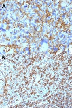
This monoclonal antibody reacts with the beta-chain of HLA-DRB1 antigen, a member of MHC class II molecules. It does not cross react with HLA-DP and HLA-DQ. HLA-DR is a heterodimeric cell surface glycoprotein comprised of a 36kDa alpha (heavy) chain and a 28kDa beta (light) chain. It is expressed on B-cells, activated T-cells, monocytes/macrophages, dendritic cells and other non-professional APCs. In conjunction with the CD3/TCR complex and CD4 molecules, HLA-DR is critical for efficient peptide presentation to CD4+ T cells. It is an excellent histiocytic marker in paraffin sections producing intense cytoplasmic staining. True histiocytic neoplasms are similarly positive. HLA-DR antigens also occur on a variety of epithelial cells and their corresponding neoplastic counterparts. Loss of HLA-DR expression is related to tumor microenvironment and predicts adverse outcome in diffuse large B-cell lymphoma.
Content And Storage
Store at 4C in the dark.
Isotype
IgG2b κ
Applications
Western Blot, Flow Cytometry, Immunohistochemistry, Immunocytochemistry, Immunofluorescence, Immunohistochemistry (Paraffin)
Clone
LN-3 + HLA-DRB/1067
Dilution
Western Blot, Flow Cytometry, Immunohistochemistry, Immunocytochemistry/Immunofluorescence, Immunohistochemistry-Paraffin, Immunofluorescence
Gene Alias
Clone P2-beta-3, DR1, DR-1, DR12, DR-12, DR13, DR-13, DR14, DR-14, DR16, DR-16, DR4, DR-4, DR5, DR-5, DR7, DR-7, DR8, DR-8, DR9, DR-9, DRB1, DRw10, DRw11, DRw8, DW2.2/DR2.2, FLJ75017, FLJ76359, HLA class II antigen beta chain, HLA class II histocompatibility antigen, DR-1 beta chain, HLA-DR1B, HLA-DRB, HLA-DRB1*, HLA-DRB2, HLA-DR-beta 1, human leucocyte antigen DRB1, leucocyte antigen DR beta 1 chain, leucocyte antigen DRB1, lymphocyte antigen DRB1, major histocompatibility complex, class II, DR beta 1, MHC class II antigen DRB1*1, MHC class II antigen DRB1*10, MHC class II antigen DRB1*11, MHC class II antigen DRB1*12, MHC class II antigen DRB1*13, MHC class II antigen DRB1*14, MHC class II antigen DRB1*15, MHC class II antigen DRB1*16, MHC class II antigen DRB1*3, MHC class II antigen DRB1*4, MHC class II antigen DRB1*7, MHC class II antigen DRB1*8, MHC class II antigen DRB1*9, MHC class II antigen HLA-DR13, MHC class II HLA-DR
Host Species
Mouse
Purification Method
Protein A or G purified
Research Discipline
Adaptive Immunity, Asthma, Cell Biology, Diabetes Research, Immunology
Gene ID (Entrez)
3123
Target Species
Human, Monkey
Form
Purified
Related Products


Description
- HLA DRB1 Monoclonal specifically detects HLA DRB1 in Human, Monkey samples
- It is validated for Western Blot, Flow Cytometry, Immunohistochemistry, Immunocytochemistry/Immunofluorescence, Immunohistochemistry-Paraffin, Immunofluorescence.
Compare Similar Items
Show Difference

Antigen: HLA DRB1
Classification: Monoclonal
Conjugate: Biotin
Formulation: PBS with 0.05% Sodium Azide
Gene Symbols: HLA-DRB1
Immunogen: Activated human peripheral blood mononuclear cells (LN-3 and HLA-DRB/1067)
Quantity: 0.1 mL
Primary or Secondary: Primary
Test Specificity: This monoclonal antibody reacts with the beta-chain of HLA-DRB1 antigen, a member of MHC class II molecules. It does not cross react with HLA-DP and HLA-DQ. HLA-DR is a heterodimeric cell surface glycoprotein comprised of a 36kDa alpha (heavy) chain and a 28kDa beta (light) chain. It is expressed on B-cells, activated T-cells, monocytes/macrophages, dendritic cells and other non-professional APCs. In conjunction with the CD3/TCR complex and CD4 molecules, HLA-DR is critical for efficient peptide presentation to CD4+ T cells. It is an excellent histiocytic marker in paraffin sections producing intense cytoplasmic staining. True histiocytic neoplasms are similarly positive. HLA-DR antigens also occur on a variety of epithelial cells and their corresponding neoplastic counterparts. Loss of HLA-DR expression is related to tumor microenvironment and predicts adverse outcome in diffuse large B-cell lymphoma.
Content And Storage: Store at 4C in the dark.
Isotype: IgG2b κ
Applications: Western Blot, Flow Cytometry, Immunohistochemistry, Immunocytochemistry, Immunofluorescence, Immunohistochemistry (Paraffin)
Clone: LN-3 + HLA-DRB/1067
Dilution: Western Blot, Flow Cytometry, Immunohistochemistry, Immunocytochemistry/Immunofluorescence, Immunohistochemistry-Paraffin, Immunofluorescence
Gene Alias: Clone P2-beta-3, DR1, DR-1, DR12, DR-12, DR13, DR-13, DR14, DR-14, DR16, DR-16, DR4, DR-4, DR5, DR-5, DR7, DR-7, DR8, DR-8, DR9, DR-9, DRB1, DRw10, DRw11, DRw8, DW2.2/DR2.2, FLJ75017, FLJ76359, HLA class II antigen beta chain, HLA class II histocompatibility antigen, DR-1 beta chain, HLA-DR1B, HLA-DRB, HLA-DRB1*, HLA-DRB2, HLA-DR-beta 1, human leucocyte antigen DRB1, leucocyte antigen DR beta 1 chain, leucocyte antigen DRB1, lymphocyte antigen DRB1, major histocompatibility complex, class II, DR beta 1, MHC class II antigen DRB1*1, MHC class II antigen DRB1*10, MHC class II antigen DRB1*11, MHC class II antigen DRB1*12, MHC class II antigen DRB1*13, MHC class II antigen DRB1*14, MHC class II antigen DRB1*15, MHC class II antigen DRB1*16, MHC class II antigen DRB1*3, MHC class II antigen DRB1*4, MHC class II antigen DRB1*7, MHC class II antigen DRB1*8, MHC class II antigen DRB1*9, MHC class II antigen HLA-DR13, MHC class II HLA-DR
Host Species: Mouse
Purification Method: Protein A or G purified
Research Discipline: Adaptive Immunity, Asthma, Cell Biology, Diabetes Research, Immunology
Gene ID (Entrez): 3123
Target Species: Human, Monkey
Form: Purified
Antigen: Mucin 5AC
Classification: Monoclonal
Conjugate: Biotin
Formulation: PBS with 0.05% Sodium Azide
Gene Symbols: MUC5AC
Immunogen: Recombinant human MUC5AC protein (MUC5AC/917); M1 mucin preparation from the fluid of an ovarian mucinous cyst belonging to an O Le(a-b) patient (45M1) (Uniprot: P98088)
Quantity: 0.1 mL
Primary or Secondary: Primary
Test Specificity: Mucin 5AC glycoprotein (MUC5AC) is a 641kDa glycoprotein belonging to the superfamily of mucins. Mucins are high molecular weight glycoproteins produced by epithelial cells and can be divided into two families; secretory mucins and membrane bound mucins. MUC5AC is a mucus-forming secreted mucin that is found in normal gastric and tracheo-bronchial mucosa, but absent from normal colon. MUC5AC expression is present in primary ovarian mucinous cancer but usually absent in colorectal adenocarcinoma, thus showing an expression pattern opposite to MUC2. Together with a panel of antibodies, Anti-MUC5AC may be useful for differential identification of primary mucinous ovarian tumors from colon adenocarcinoma metastatic to the ovary. MUC5AC antibodies may also be useful for identification of intestinal metaplasia as well as in the identification of pancreatic carcinoma and pre-cancerous changes vs. normal pancreas.
Content And Storage: Store at 4C in the dark.
Isotype: IgG1 κ
Applications: Flow Cytometry, Immunohistochemistry, Immunohistochemistry (Paraffin), Immunofluorescence
Clone: MUC5AC/917 + 45M1
Dilution: Flow Cytometry, Immunohistochemistry, Immunohistochemistry-Paraffin, Immunofluorescence
Gene Alias: gastric mucin, leB, lewis B blood group antigen, major airway glycoprotein, MUC5, mucin 5, subtypes A and C, tracheobronchial/gastric, mucin 5AC, oligomeric mucus/gel-forming, mucin 5AC, oligomeric mucus/gel-forming pseudogene, mucin-5 subtype AC, tracheobronchial, mucin-5AC, TBM, tracheobronchial mucin
Host Species: Mouse
Purification Method: Protein A or G purified
Research Discipline: Cellular Markers, Extracellular Matrix, Infections (Virus Bacteria and Parasites), Signal Transduction
Gene ID (Entrez): 4586
Target Species: Human
Form: Purified
Antigen: c-Myc
Classification: Monoclonal
Conjugate: Biotin
Formulation: PBS with 0.05% Sodium Azide
Gene Symbols: MYC
Immunogen: Recombinant human c-Myc protein (Uniprot: P01106)
Quantity: 0.1 mL
Primary or Secondary: Primary
Test Specificity: It recognizes a transcription factor of 64-67kDa, identified as c-myc. This monoclonal antibody shows no cross-reaction with v-myc. c-myc is involved in the control of cell proliferation and differentiation and is amplified and/or over-expressed in a variety of tumors. Over-expression of c-myc protein occurs frequently in luminal cells of prostate intraepithelial neoplasia as well as in most primary carcinomas and metastatic disease. Rearrangement of the MYC gene is found in 3% to 16% of diffuse large B-cell lymphoma (DLBCLs) and in nearly 100% of Burkitt lymphomas (BL). Identifying MYC status is important in establishing final diagnosis of DLBCL, BL, or B-cell lymphoma, with features intermediate between DLBCL and BL as well as in differential diagnoses of the lymphomas.
Content And Storage: Store at 4C in the dark.
Isotype: IgG1 κ
Applications: Flow Cytometry, Immunohistochemistry, Immunohistochemistry (Paraffin), Immunofluorescence
Clone: MYC275 + MYC909
Dilution: Flow Cytometry, Immunohistochemistry, Immunohistochemistry-Paraffin, Immunofluorescence
Gene Alias: avian myelocytomatosis viral oncogene homolog, BHLHE39, bHLHe39MRTL, Class E basic helix-loop-helix protein 39, c-Myc, MYC, myc proto-oncogene protein, MYCC, myc-related translation/localization regulatory factor, Proto-oncogene c-Myc, Transcription factor p64, v-myc avian myelocytomatosis viral oncogene homolog, v-myc myelocytomatosis viral oncogene homolog (avian)
Host Species: Mouse
Purification Method: Protein A or G purified
Research Discipline: Autophagy, Cancer, Cancer Stem Cells, Cell Cycle and Replication, Chromatin Research, Core ESC Like Genes, Epigenetics, Epitope Tags, Stem Cell Markers, Transcription Factors and Regulators, Tumor Suppressors
Gene ID (Entrez): 4609
Target Species: Human
Form: Purified