50-113-7621
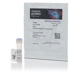
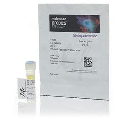
Molecular Probes™ Vybrant™ DyeCycle™ Violet Stain
Manufacturer: Molecular Probes™
Select a Size
| Pack Size | SKU | Availability | Price |
|---|---|---|---|
| Each of 1 | 50-113-7621-Each-of-1 | In Stock | ₹ 50,018.00 |
50-113-7621 - Each of 1
In Stock
Quantity
1
Base Price: ₹ 50,018.00
GST (18%): ₹ 9,003.24
Total Price: ₹ 59,021.24
Concentration
5 mM
Detection Method
Fluorescence
Product Type
Stain
Emission
369⁄437
Format
Tube(s)
Quantity
200 Reactions
Solubility
DMSO (Dimethylsulfoxide)
Content And Storage
Contains 1 vial of Vybrant™ DyeCycle™ violet stain (5 mM in DMSO).Store in refrigerator (2°C to 8°C) and protect from light.
For Use With (Equipment)
Flow Cytometer
Dye Type
Vybrant™ DyeCycle™ Violet
Form
Solution
Product Line
DyeCycle™
Shipping Condition
Room Temperature
Sub Cellular Localization
Nucleic Acids
Description
- Vybrant™ DyeCycle™ Violet Stain is a cell permeable DNA dye that can be used for cell cycle analysis and stem cell side population by flow cytometry
- Precise—accurate cell cycle analysis in living cells Safe—low cytotoxicity for cell sorting and additional live cell experiments Minimal compensation—easier multiplexing Flexible Simple, robust staining protocol View a selection guide for all products related to cell cycle analysis of fixed and live cells in flow cytometry
- Precise Successful cell cycle analysis requires a dye that is DNA selective and can stain cells in a homogeneous pattern minimizing fluorescence variability
- The Vybrant™ DyeCycle™ Violet Stain is an ideal tool for DNA content analysis in living cells since the stain is cell permeable and, after binding double-stranded DNA, emits a fluorescent signal that is proportional to the DNA mass (see figure)
- Low Cytotoxicity Unlike stains that require high concentrations or have chemical structures that are toxic to cells, Vybrant™ DyeCycle™ stains exhibit relatively low cytotoxicity, allowing the possibility of sorting based on the phase of the cell cycle
- Minimal Compensation Well-suited for the popular violet laser line, the Vybrant™ DyeCycle™ Violet Stain/DNA complex has fluorescence excitation and emission maxima of 369/437 nm, respectively (see figure)
- The violet excitation and narrow emission of Vybrant™ DyeCycle™ Violet Stain make it ideal for multiplexing due to the limited spectral overlap with other common dyes (Alexa Fluor™ 488, FITC, and RPE) and fluorescent proteins (Green Fluorescent Protein (GFP) and mCherry)
- Vybrant™ DyeCycle™ Violet Stain can also be used with UV excitation, having emission at ∼440 nm
- Flexible Vybrant™ DyeCycle™ Violet Stain has been shown to not only work for both live cells and fixed cells in cell cycle assays, but to identify stem cell side populations and early progenitors in mammalian hematopoietic tissues (see figure)
- Simple, Robust Staining Protocol For cell analysis, simply prepare flow cytometry tubes each containing 1 mL of cells suspended in complete media at a cell concentration of 1 × 10 6 cells/mL
- To each tube, add 1μL of Vybrant™ DyeCycle™ Violet Stain and mix well
- Final stain concentration is 5μM
- Incubate at 37°C for 30 minutes, protected from light
- Keep cells at 37°C until acquisition
- Analyze samples without washing or fixing on a flow cytometer using ∼405 nm excitation and ∼440 nm emission.
Compare Similar Items
Show Difference
Concentration: 5 mM
Detection Method: Fluorescence
Product Type: Stain
Emission: 369⁄437
Format: Tube(s)
Quantity: 200 Reactions
Solubility: DMSO (Dimethylsulfoxide)
Content And Storage: Contains 1 vial of Vybrant™ DyeCycle™ violet stain (5 mM in DMSO).Store in refrigerator (2°C to 8°C) and protect from light.
For Use With (Equipment): Flow Cytometer
Dye Type: Vybrant™ DyeCycle™ Violet
Form: Solution
Product Line: DyeCycle™
Shipping Condition: Room Temperature
Sub Cellular Localization: Nucleic Acids
Concentration:
5 mM
Detection Method:
Fluorescence
Product Type:
Stain
Emission:
369⁄437
Format:
Tube(s)
Quantity:
200 Reactions
Solubility:
DMSO (Dimethylsulfoxide)
Content And Storage:
Contains 1 vial of Vybrant™ DyeCycle™ violet stain (5 mM in DMSO).Store in refrigerator (2°C to 8°C) and protect from light.
For Use With (Equipment):
Flow Cytometer
Dye Type:
Vybrant™ DyeCycle™ Violet
Form:
Solution
Product Line:
DyeCycle™
Shipping Condition:
Room Temperature
Sub Cellular Localization:
Nucleic Acids
Concentration: __
Detection Method: __
Product Type: Dead Cell Apoptosis Kit
Emission: __
Format: Tube, Slide
Quantity: __
Solubility: __
Content And Storage: Contains 1 vial of PO-PRO™-1 dye (500 μL), and 1 vial of 7-AAD (7-aminoactinomycin D, 200 μL).Store in refrigerator (2–8°C) and protect from light.
For Use With (Equipment): Fluorescence Microscope, Flow Cytometer
Dye Type: __
Form: __
Product Line: Vybrant™
Shipping Condition: Room Temperature
Sub Cellular Localization: __
Concentration:
__
Detection Method:
__
Product Type:
Dead Cell Apoptosis Kit
Emission:
__
Format:
Tube, Slide
Quantity:
__
Solubility:
__
Content And Storage:
Contains 1 vial of PO-PRO™-1 dye (500 μL), and 1 vial of 7-AAD (7-aminoactinomycin D, 200 μL).Store in refrigerator (2–8°C) and protect from light.
For Use With (Equipment):
Fluorescence Microscope, Flow Cytometer
Dye Type:
__
Form:
__
Product Line:
Vybrant™
Shipping Condition:
Room Temperature
Sub Cellular Localization:
__
Concentration: __
Detection Method: __
Product Type: __
Emission: __
Format: __
Quantity: 25 Reactions
Solubility: __
Content And Storage: Contains 1 vial of Zenon APC mouse IgG2alabeling reagent (125 μL), and 1 vial of Zenon blocking reagent (mouse IgG, 125 μL).Store in refrigerator (2–8°C) and protect from light.
For Use With (Equipment): __
Dye Type: __
Form: __
Product Line: Zenon™
Shipping Condition: __
Sub Cellular Localization: __
Concentration:
__
Detection Method:
__
Product Type:
__
Emission:
__
Format:
__
Quantity:
25 Reactions
Solubility:
__
Content And Storage:
Contains 1 vial of Zenon APC mouse IgG2alabeling reagent (125 μL), and 1 vial of Zenon blocking reagent (mouse IgG, 125 μL).Store in refrigerator (2–8°C) and protect from light.
For Use With (Equipment):
__
Dye Type:
__
Form:
__
Product Line:
Zenon™
Shipping Condition:
__
Sub Cellular Localization:
__
Concentration: __
Detection Method: __
Product Type: __
Emission: __
Format: __
Quantity: 25 Reactions
Solubility: __
Content And Storage: Contains 1 vial of Zenon APC Human IgG labeling reagent (125 μL), and 1 vial of Zenon blocking reagent (human IgG, 125 μL).Store in refrigerator (2–8°C) and protect from light.
For Use With (Equipment): __
Dye Type: __
Form: __
Product Line: Zenon™
Shipping Condition: __
Sub Cellular Localization: __
Concentration:
__
Detection Method:
__
Product Type:
__
Emission:
__
Format:
__
Quantity:
25 Reactions
Solubility:
__
Content And Storage:
Contains 1 vial of Zenon APC Human IgG labeling reagent (125 μL), and 1 vial of Zenon blocking reagent (human IgG, 125 μL).Store in refrigerator (2–8°C) and protect from light.
For Use With (Equipment):
__
Dye Type:
__
Form:
__
Product Line:
Zenon™
Shipping Condition:
__
Sub Cellular Localization:
__
