D23841
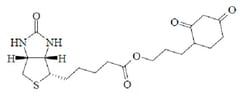
Invitrogen™ DAF-FM (4-Amino-5-Methylamino-2',7'-Difluorofluorescein)
Manufacturer: Invitrogen™
Select a Size
| Pack Size | SKU | Availability | Price |
|---|---|---|---|
| Each of 1 | D23841-Each-of-1 | In Stock | ₹ 70,310.00 |
D23841 - Each of 1
In Stock
Quantity
1
Base Price: ₹ 70,310.00
GST (18%): ₹ 12,655.80
Total Price: ₹ 82,965.80
Quantity
1 mg
Related Products


Description
- DAF-FM is essentially nonfluorescent until it reacts with NO to form fluorescent benzotriazole
- DAF-FM fluorescence can be detected by any instrument that can detect fluorescein, including flow cytometers, microscopes, fluorescent microplate readers and fluorometers
- Ex/Em of DAF-FM: approx
- 495/515nm Lyophilized product should be dissolved using DMSO and then added to an aqueous buffer to create a working solution DAF-FM can be introduced into cells by pressure injection or perfusion from patch-clamp pipette Buffers containing bovine serum albumin (BSA) and phenol red may affect the fluorescence and should be used with caution Fluorescence quantum yield of DAF-FM is ∽0.005, but increases about 160-fold, to ∽0.81, after reacting with NO Assessment of NO production in transaldolase-deficient lymphoblasts by flow cytometry Detection of NO accumulation in embryonic cortical neurons following neurotrophin stimulation in vivo imaging of NO in zebrafish Intravital microscopic detection of NO generation associated with angiogenesis in mice Quantitation of ATP-induced NO release in rabbit platelets Spectra of NO adduct of DAF-FM are independent of pH above pH 5.5 NO adduct of DAF-FM is significantly more photostable than that of DAF-2, which means additional time for image capture DAF-FM is more sensitive reagent for NO than is DAF-2 (NO detection limit for DAF-FM approx
- 3nM versus approx
- 5nM for DAF-2) Refer to the Molecular Probes™ Handbook for additional product information
- Anion Detection, Cell Analysis, Cell Metabolism, Cell Viability, Proliferation and Function, Ionic Homeostasis and Signaling, Nitric Oxide Research, Nitro-Oxidative Stress Order Info Shipping Condition: Room Temperature