05-373
Anti-Cyclin B1 Antibody, clone GNS3 (8A5D12)
clone GNS3 (8A5D12), Upstate®, from mouse
Manufacturer: Sigma Aldrich
Synonym(S): G2/mitotic-specific cyclin B1, cyclin B1
Select a Size
| Pack Size | SKU | Availability | Price |
|---|---|---|---|
| 25 μG | 05-373-25-μG | In Stock | ₹ 13,730.00 |
| 200 μG | 05-373-200-μG | In Stock | ₹ 42,460.00 |
05-373 - 25 μG
In Stock
Quantity
1
Base Price: ₹ 13,730.00
GST (18%): ₹ 2,471.40
Total Price: ₹ 16,201.40
biological source
mouse
Quality Level
100
antibody form
purified antibody
antibody product type
primary antibodies
clone
GNS3 (8A5D12), monoclonal
species reactivity
human, mouse
packaging
antibody small pack of 25 μg
manufacturer/tradename
Upstate®
technique(s)
immunohistochemistry: suitableimmunoprecipitation (IP): suitablewestern blot: suitable
isotype
IgG
Description
- General description: G2/mitotic-specific cyclin-B1 (UniProt: P14635; also known as Cyclin B1) is encoded by the CCNB1 (also known as CCNB) gene (Gene ID: 891) in human. Cyclins are the regulatory subunits of the cell cycle-dependent kinases (CDKs) that are responsible for the phosphorylation of several cellular targets. Cyclins contain the nuclear localization sequence (NLS) that help move CDKs into the nucleus. They also contain PEST (Pro, Glu, Ser, and Thr) sequences that target them for degradation by the ubiquitin-proteasomal pathway. Once the CDKs have completed their role, they undergo a rapid programmed proteolysis via ubiquitin-mediated delivery to the proteasome complex. Cyclin B1, a regulatory protein involved in mitosis, complexes with CDK1 to form the maturation-promoting factor (MPF). It is shown to be essential for the control of the cell cycle at the G2/M (mitosis) transition. It accumulates steadily during G2 phase and is abruptly destroyed at mitosis. Hence, the cyclin B1-CDK1 complex is considered to be a key regulator for mitotic entry. This complex phosphorylates a number of proteins prior to mitotic entry. Although five serine phosphorylation sites are described for cyclin B1, (Ser 116, 126, 128, 133, and 147), serine 133 phosphorylation by PLK1 regulates the entry of Cyclin B1-CDK1 complex into the nucleus during prophase. At the end of mitosis, cyclin B1 is rapidly removed by a ubiquitin ligase (anaphase-promoting complex/cyclosome) loaded with the targeting subunit CDC20. Activated cyclin B1-CDK1 complex is reported to catalyze its own destruction by stimulating the activity of APC. (Ref.: Van Zon, W., et al. (2010). J. Cell. Biol. 190(4); 587-602; Yuan, J., et al. (2004). Oncogene 23(34); 5843-5852).
- Specificity: In addition to human, weak species cross-reactivity was observed with mouse.
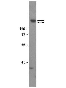
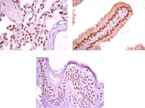
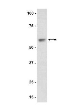
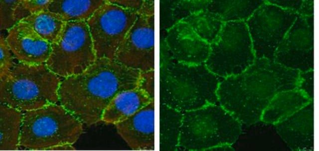
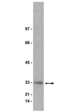
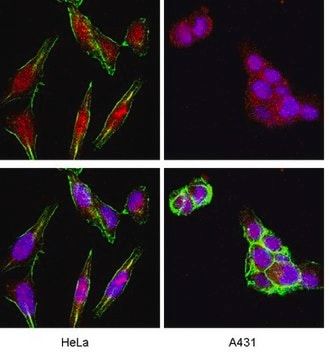
- Application: Immunoprecipitation: 2 μg of a previous lot immunoprecipitated human cyclin B1 and cdc2 kinase from 500 μg of A431 RIPA lysate as determined by a subsequent immunoblot of the immunoprecipitate using anti-cdk1/cdc2 (PSTAIR), (Catalog # 06-923).Western Blotting Analysis: 0.1 mg/mL of this antibody detected Cyclin B1 in A431 cell lysate.Immunohistochemistry (Paraffin) Analysis: A 1:250 and 1:50 dilutions of this antibody detected Cyclin B1 in Human tonsil and Human bone marrow tissue sections, respectively.
- Quality: Routinely evaluated by western blot analysis on RIPA lysate from human A431 carcinoma cells.Western Blot Analysis: 0.5-2 μg/mL of this lot detected cyclin B1 in RIPA lysates from human A431 carcinoma cells.
- Target description: 58 kDa
- Linkage: Replaces: 04-220
- Physical form: Format: Purified
- Analysis Note: ControlPositive Antigen Control: Catalog #12-301, non-stimulated A431 cell lysate. Add 2.5µL of 2-mercaptoethanol/100µL of lysate and boil for 5 minutes to reduce the preparation. Load 20µg of reduced lysate per lane for minigels.
- Other Notes: Concentration: Please refer to the Certificate of Analysis for the lot-specific concentration.
- Legal Information: UPSTATE is a registered trademark of Merck KGaA, Darmstadt, Germany
Compare Similar Items
Show Difference
biological source: mouse
Quality Level: 100
antibody form: purified antibody
antibody product type: primary antibodies
clone: GNS3 (8A5D12), monoclonal
species reactivity: human, mouse
packaging: antibody small pack of 25 μg
manufacturer/tradename: Upstate®
technique(s): immunohistochemistry: suitableimmunoprecipitation (IP): suitablewestern blot: suitable
isotype: IgG
biological source:
mouse
Quality Level:
100
antibody form:
purified antibody
antibody product type:
primary antibodies
clone:
GNS3 (8A5D12), monoclonal
species reactivity:
human, mouse
packaging:
antibody small pack of 25 μg
manufacturer/tradename:
Upstate®
technique(s):
immunohistochemistry: suitableimmunoprecipitation (IP): suitablewestern blot: suitable
isotype:
IgG
