MAB3420
Anti-Tau-1 Antibody, clone PC1C6
clone PC1C6, Chemicon®, from mouse
Manufacturer: Sigma Aldrich
Synonym(S): Anti-Tau Antibody
Select a Size
| Pack Size | SKU | Availability | Price |
|---|---|---|---|
| 25 μG | MAB3420-25-μG | In Stock | ₹ 13,730.00 |
| 100 μG | MAB3420-100-μG | In Stock | ₹ 63,150.00 |
MAB3420 - 25 μG
In Stock
Quantity
1
Base Price: ₹ 13,730.00
GST (18%): ₹ 2,471.40
Total Price: ₹ 16,201.40
biological source
mouse
Quality Level
100
antibody form
purified antibody
clone
PC1C6, monoclonal
species reactivity
human, rat, bovine
packaging
antibody small pack of 25 μg
manufacturer/tradename
Chemicon®
technique(s)
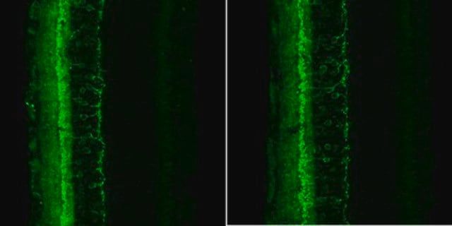
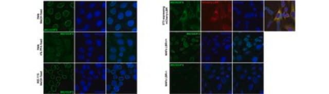
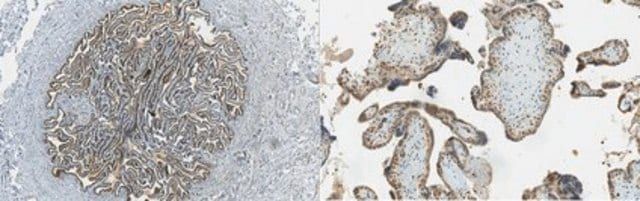
immunofluorescence: suitableimmunohistochemistry: suitablewestern blot: suitable
isotype
IgG2a
NCBI accession no.
NM_005910.3NM_016834.2NM_016835.2NM_016841.2
Related Products


Description
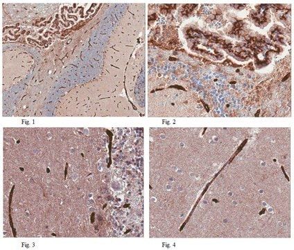
- General description: Tau, a microtubulebinding protein which serves to stabilize microtubules in growing axons, is found to be hyperphosphorylated in paired helical filaments (PHF), the major fibrous component of neurofibrillary lesions associated with Alzheimer’s disease. Hyperphosphorylation of Tau is thought to be the critical event leading to the assembly of PHF. Six Tau protein isoforms have been identified, all of which are phosphorylated by glycogen synthase kinase 3 (GSK 3). Cellular and subcellular localization: In situ, anti-tau-1 has a stringent specificity for the axons of neurons. The antibody does not stain the cell bodies or dendrites of neurons, nor does it stain any other cell type (4). However, this in vivo intracellular specificity is not maintained in culture: anti-tau-1 stains the axon, cell bodies, and dendrites of rat hippocampal neurons grown in culture (5). The specificity of anti-tau-1 was originally thought to represent the restricted expression of tau to axons. Later studies revealed that this specificity is dependant on the state of phosphorylation. In dephosphorylated samples (samples treated with alkaline phosphatase) anti-tau-1 stains astrocytes, perineuronal glial cells, and the axons, cell bodies and dendrites of neurons, while in untreated samples, anti-tau-1 stains only axons (6). (The epitope recognized by anti-tau-1 is probably at or near a phosphorylated site.)
- Specificity: Anti-Tau-1 Antibody, clone PC1C6 binds to all known electrophoretic species of tau in human, rat and bovine brain (one-dimensional SDS-PAGE). However there is some unphosphorylated bias with clone PC1C6 as it seem to recognize only dephosphorylated serine sites at 195, 198, 199, and 202 {Szendrei, et al 1993; http://www.ncbi.nlm.nih.gov/entrez/query.fcgi-cmd=Retrieve&db=pubmed&dopt=Abstract&list_uids=7680727}. Also see Billingsley & Kincaid, 1997 Biochem J 323:577-591 for additional mapping information on PC1C6.
- Immunogen: Purified denatured bovine microtubule associated proteins.
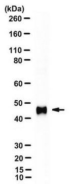
- Application: Anti-Tau-1 Antibody, clone PC1C6 is an antibody against Tau-1 for use in IH & WB with more than 65 product citations.
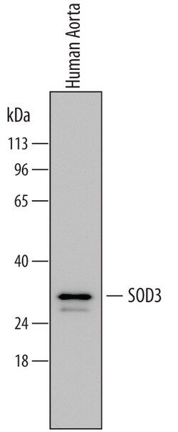
- Target description: 5 bands (52–68 kDa)
- Linkage: Replaces: AB1512
- Physical form: 0.02M phosphate buffer, pH 7.6, 0.25M NaCl, and 0.1% sodium azide
- Storage and Stability: Maintain for 1 year at -20°C from date of shipment. Aliquot to avoid repeated freezing and thawing. For maximum recovery of product, centrifuge the original vial after thawing and prior to removing the cap.
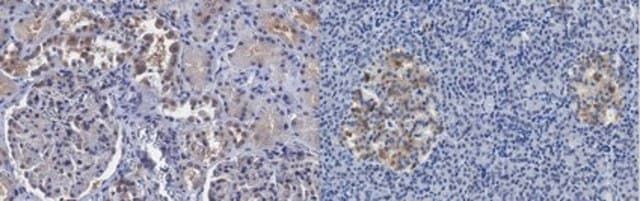
- Analysis Note: ControlAlzheimer′s brain tissue (dephosphorylation with alkaline phosphatase is recommended for staining neurofibrillary tangles in Alzheimer’s brain tissue) or human T98G glioblastoma cells
- Other Notes: Concentration: Please refer to the Certificate of Analysis for the lot-specific concentration.
- Legal Information: CHEMICON is a registered trademark of Merck KGaA, Darmstadt, Germany
- Disclaimer: Unless otherwise stated in our catalog or other company documentation accompanying the product(s), our products are intended for research use only and are not to be used for any other purpose, which includes but is not limited to, unauthorized commercial uses, in vitro diagnostic uses, ex vivo or in vivo therapeutic uses or any type of consumption or application to humans or animals.
SAFETY INFORMATION
WGK
WGK 2
Flash Point(F)
Not applicable
Flash Point(C)
Not applicable
Compare Similar Items
Show Difference

biological source: mouse
Quality Level: 100
antibody form: purified antibody
clone: PC1C6, monoclonal
species reactivity: human, rat, bovine
packaging: antibody small pack of 25 μg
manufacturer/tradename: Chemicon®
technique(s): immunofluorescence: suitableimmunohistochemistry: suitablewestern blot: suitable
isotype: IgG2a
NCBI accession no.: NM_005910.3NM_016834.2NM_016835.2NM_016841.2
biological source:
mouse
Quality Level:
100
antibody form:
purified antibody
clone:
PC1C6, monoclonal
species reactivity:
human, rat, bovine
packaging:
antibody small pack of 25 μg
manufacturer/tradename:
Chemicon®
technique(s):
immunofluorescence: suitableimmunohistochemistry: suitablewestern blot: suitable
isotype:
IgG2a
NCBI accession no.:
NM_005910.3NM_016834.2NM_016835.2NM_016841.2
biological source: __
Quality Level: __
antibody form: __
clone: __
species reactivity: __
packaging: __
manufacturer/tradename: __
technique(s): __
isotype: __
NCBI accession no.: __
biological source:
__
Quality Level:
__
antibody form:
__
clone:
__
species reactivity:
__
packaging:
__
manufacturer/tradename:
__
technique(s):
__
isotype:
__
NCBI accession no.:
__