HSP60 Antibody (HSPD1/875), Novus Biologicals™
Mouse Monoclonal Antibody
Manufacturer: Fischer Scientific
The price for this product is unavailable. Please request a quote
Antigen
HSP60
Concentration
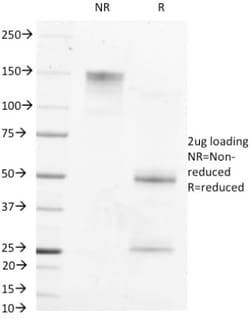
0.2 mg/mL
Applications
Flow Cytometry, Immunofluorescence
Conjugate
Unconjugated
Host Species
Mouse
Research Discipline
Apoptosis, Cellular Markers, Membrane Trafficking and Chaperones, Mitochondrial Markers
Formulation
10mM PBS and 0.05% BSA with 0.05% Sodium Azide
Gene Alias
60 kDa chaperonin, Chaperonin 60, CPN60, GROEL, heat shock 60kD protein 1 (chaperonin), heat shock 60kDa protein 1 (chaperonin), Heat shock protein 60, heat shock protein 65, HLD4, Hsp60, HSP-60, HSP60SPG13, HSP65, HuCHA60, Mitochondrial matrix protein P1,60 kDa heat shock protein, mitochondrial, P60 lymphocyte protein, short heat shock protein 60 Hsp60s1, spastic paraplegia 13 (autosomal dominant)
Gene Symbols
HSPD1
Isotype
IgG1 κ
Purification Method
Protein A or G purified
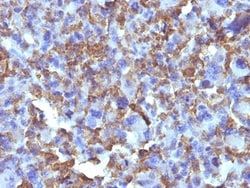
Test Specificity
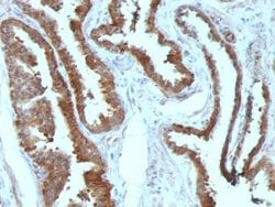
Recognizes a 60kDa protein, identified as the heat shock protein 60 (hsp60). A wide variety of environmental and pathophysiological stressful conditions trigger the synthesis of a family of proteins known as heat shock proteins (hsps), more appropriately called as stress response proteins (srps). hsp60 is a potential antigen in a number of autoimmune diseases. In human arthritis and in experimentally induced arthritis in animals, disease development coincides with the development of immune reactivity directed against not only bacterial hsp60, but also against its mammalian homolog.
Clone
HSPD1/875
Dilution
Flow Cytometry 0.5 - 1 ug/million cells in 0.1 ml, Immunofluorescence 0.5 - 1.0 ug/ml
Classification
Monoclonal
Form
Purified
Regulatory Status
RUO
Target Species
Human, Mouse, Rat, Chicken, Guinea Pig, Hamster, Monkey
Gene Accession No.
P10809
Gene ID (Entrez)
3329
Immunogen
Recombinant human HSPD1 protein
Primary or Secondary
Primary
Content And Storage
Store at 4C.
Molecular Weight of Antigen
60 kDa
Related Products

Description
- Ensure accurate, reproducible results in Flow Cytometry, Immunofluorescence HSP60 Monoclonal specifically detects HSP60 in Human, Mouse, Rat, Chicken, Guinea Pig, Hamster, Monkey samples
- It is validated for Western Blot, Immunocytochemistry/Immunofluorescence, Immunohistochemistry-Paraffin, Protein Array.
Compare Similar Items
Show Difference
Antigen: HSP60
Concentration: 0.2 mg/mL
Applications: Flow Cytometry, Immunofluorescence
Conjugate: Unconjugated
Host Species: Mouse
Research Discipline: Apoptosis, Cellular Markers, Membrane Trafficking and Chaperones, Mitochondrial Markers
Formulation: 10mM PBS and 0.05% BSA with 0.05% Sodium Azide
Gene Alias: 60 kDa chaperonin, Chaperonin 60, CPN60, GROEL, heat shock 60kD protein 1 (chaperonin), heat shock 60kDa protein 1 (chaperonin), Heat shock protein 60, heat shock protein 65, HLD4, Hsp60, HSP-60, HSP60SPG13, HSP65, HuCHA60, Mitochondrial matrix protein P1,60 kDa heat shock protein, mitochondrial, P60 lymphocyte protein, short heat shock protein 60 Hsp60s1, spastic paraplegia 13 (autosomal dominant)
Gene Symbols: HSPD1
Isotype: IgG1 κ
Purification Method: Protein A or G purified
Test Specificity: Recognizes a 60kDa protein, identified as the heat shock protein 60 (hsp60). A wide variety of environmental and pathophysiological stressful conditions trigger the synthesis of a family of proteins known as heat shock proteins (hsps), more appropriately called as stress response proteins (srps). hsp60 is a potential antigen in a number of autoimmune diseases. In human arthritis and in experimentally induced arthritis in animals, disease development coincides with the development of immune reactivity directed against not only bacterial hsp60, but also against its mammalian homolog.
Clone: HSPD1/875
Dilution: Flow Cytometry 0.5 - 1 ug/million cells in 0.1 ml, Immunofluorescence 0.5 - 1.0 ug/ml
Classification: Monoclonal
Form: Purified
Regulatory Status: RUO
Target Species: Human, Mouse, Rat, Chicken, Guinea Pig, Hamster, Monkey
Gene Accession No.: P10809
Gene ID (Entrez): 3329
Immunogen: Recombinant human HSPD1 protein
Primary or Secondary: Primary
Content And Storage: Store at 4C.
Molecular Weight of Antigen: 60 kDa
Antigen: Macrophage and Histiocytoma Marker
Concentration: 0.2mg/mL
Applications: Immunohistochemistry (Paraffin)
Conjugate: Unconjugated
Host Species: Mouse
Research Discipline: __
Formulation: 10mM PBS with 0.05% Sodium Azide
Gene Alias: __
Gene Symbols: __
Isotype: IgG1 κ
Purification Method: Protein A or G purified
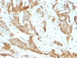
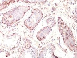
Test Specificity: In Western blotting, it detects an antigen of 125kDa in human liver and 135kDa in tumors of histiocytic origin. Comparative study of this MAb and a standard CD68 MAb showed that their antigens are different. Its antigen in all macrophage types studied is located on the plasma membrane and within cytoplasmic structures including lysosomes. This MAb shows a restricted reactivity to cells of the monocyte/macrophage system. It specifically reacts with blood monocytes and stains resident macrophages in a wide variety of human tissues. This MAb does not stain antigen-presenting cells, e.g., Langerhans cells. Reportedly, its reactivity is restricted to histiocytes and macrophages.
Clone: D11
Dilution: Immunohistochemistry-Paraffin 0.5 - 1.0 ug/ml
Classification: Monoclonal
Form: Purified
Regulatory Status: RUO
Target Species: Human, Mouse (Negative), Porcine (Negative), Rat (Negative)
Gene Accession No.: __
Gene ID (Entrez): __
Immunogen: Membrane preparation from human hepatocytes
Primary or Secondary: Primary
Content And Storage: Store at 4C.
Molecular Weight of Antigen: __
Antigen: MAGE 1
Concentration: 0.2mg/mL
Applications: Flow Cytometry, Immunohistochemistry (Paraffin), Immunofluorescence
Conjugate: Unconjugated
Host Species: Mouse
Research Discipline: Apoptosis, Cancer, Melanoma Cell Markers, Tumor Suppressors
Formulation: 10mM PBS and 0.05% BSA with 0.05% Sodium Azide
Gene Alias: Antigen MZ2-E, Cancer/testis antigen 1.1, cancer/testis antigen family 1, member 1, CT1.1melanoma antigen family A 1, MAGE-1 antigen, MAGE1A, MAGE1melanoma antigen MAGE-1, melanoma antigen family A, 1 (directs expression of antigen MZ2-E), melanoma-associated antigen 1, melanoma-associated antigen MZ2-E, MGC9326
Gene Symbols: MAGEA1
Isotype: IgG1 κ
Purification Method: Protein A or G purified
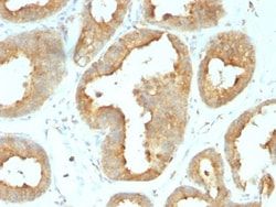
Test Specificity: Recognizes a protein of 42-46kDa, identified as MAGE-1. This MAb does not cross-react with other members of MAGE-family. Human malignant neoplasms carry rejection antigens that are recognized by the patients' autologous, tumor directed and specific, cytolytic, CD8+ T lymphocyte clones (CTL). The MAGE family of genes codes an important group of antigens. It was identified that melanomas and primary glial brain tumors express common melanoma associated antigens (MAAs). Because MAGE-1 is expressed on a significant proportion of human neoplasms of various histological types (melanoma, brain tumors of glial origin, neuroblastoma, non-small cell lung cancer, breast, gastric, colorectal, ovarian, renal cell carcinomas) and not on normal tissues, the encoded antigen may serve as a marker of early detection and target for cancer immunotherapy.
Clone: MZ2E/838
Dilution: Flow Cytometry 0.5 - 1 ug/million cells in 0.1 ml, Immunohistochemistry-Paraffin 0.5 - 1.0 ug/ml, Immunofluorescence 1 - 2 ug/ml
Classification: Monoclonal
Form: Purified
Regulatory Status: RUO
Target Species: Human
Gene Accession No.: __
Gene ID (Entrez): 4100
Immunogen: Recombinant human MAGEA1 protein
Primary or Secondary: Primary
Content And Storage: Store at 4C.
Molecular Weight of Antigen: __