MEKK1 Antibody (2F6), Novus Biologicals™
Mouse Monoclonal Antibody has been used in 1 publication
Manufacturer: Fischer Scientific
The price for this product is unavailable. Please request a quote
Antigen
MEKK1
Concentration
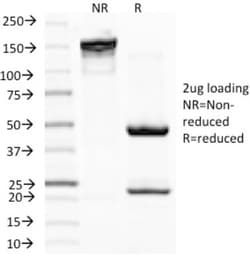
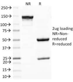
0.2mg/mL
Applications
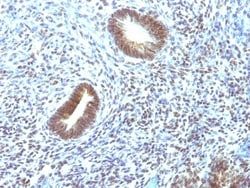
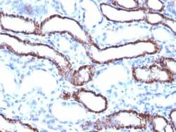
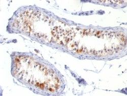
Flow Cytometry, Immunohistochemistry (Paraffin), Immunofluorescence
Conjugate
Unconjugated
Host Species
Mouse
Research Discipline
Angiogenesis, Breast Cancer, Cancer, Lipid and Metabolism, MAP Kinase Signaling, mTOR Pathway, Protein Kinase, Signal Transduction, Tyrosine Kinases
Formulation
10mM PBS and 0.05% BSA with 0.05% Sodium Azide
Gene Alias
MAPK/ERK kinase kinase 1, MAPKKK1MAP/ERK kinase kinase 1, MEK kinase 1, MEKK1EC 2.7.11.25, MEKKMEKK 1, mitogen-activated protein kinase kinase kinase 1
Gene Symbols
MAP3K1
Isotype
IgG2a κ
Purification Method
Protein A or G purified
Test Specificity
Mitogen-activated protein (MAP) kinase cascades are activated by various extracellular stimuli, including growth factors. The MEK kinases (also designated MAP kinase kinase kinases, MKKKs, MAP3Ks or MEKKs) phosphorylate and thereby activate the MEKs (also called MAP kinase kinases or MKKs), including ERK, JNK and p38. These activated MEKs in turn phosphorylate and activate the MAP kinases. The MEK kinases include Raf-1, Raf-B, Mos, MEK kinase-1, MEK kinase-2, MEK kinase-3, MEK kinase-4 and ASK 1 (MEK kinase- 5). MEK kinase-1 activates the ERK and c-Jun NH2-terminal kinase (JNK) pathways by phosphorylation of MAP2K1 and MAP2K4, and also activates the central protein kinases of the NF B pathway, CHUK and IKBKB. Additionally, MEK kinase-1 uses an E3 ligase through its PHD domain, a RING-finger-like structure, to target proteins for degradation through ubiquitination.
Clone
2F6
Dilution
Flow Cytometry 0.5 - 1 ug/million cells in 0.1 ml, Immunohistochemistry-Paraffin 0.5 - 1.0 ug/ml, Immunofluorescence 1 - 2 ug/ml
Classification
Monoclonal
Form
Purified
Regulatory Status
RUO
Target Species
Human
Gene Accession No.
Q13233
Gene ID (Entrez)
4214
Immunogen
Partial recombinant MAP3K1 (aa1211-1310) (SKNSMTLDLNSSSKCDDSFGCSSNSSNAVIPSDETVFTP-VEEKCRLDVNTELNSSIEDLLEASMPSSDTTVTFKSEVAVLSPEKAENDDTYKDDVNHNQK)
Primary or Secondary
Primary
Content And Storage
Store at 4C.
Related Products

Description
- Ensure accurate, reproducible results in Western Blot, Flow Cytometry, Immunohistochemistry (Paraffin), Immunofluorescence MEKK1 Monoclonal specifically detects MEKK1 in Human samples
- It is validated for Western Blot, Immunohistochemistry, Immunohistochemistry-Paraffin.
Compare Similar Items
Show Difference
Antigen: MEKK1
Concentration: 0.2mg/mL
Applications: Flow Cytometry, Immunohistochemistry (Paraffin), Immunofluorescence
Conjugate: Unconjugated
Host Species: Mouse
Research Discipline: Angiogenesis, Breast Cancer, Cancer, Lipid and Metabolism, MAP Kinase Signaling, mTOR Pathway, Protein Kinase, Signal Transduction, Tyrosine Kinases
Formulation: 10mM PBS and 0.05% BSA with 0.05% Sodium Azide
Gene Alias: MAPK/ERK kinase kinase 1, MAPKKK1MAP/ERK kinase kinase 1, MEK kinase 1, MEKK1EC 2.7.11.25, MEKKMEKK 1, mitogen-activated protein kinase kinase kinase 1
Gene Symbols: MAP3K1
Isotype: IgG2a κ
Purification Method: Protein A or G purified
Test Specificity: Mitogen-activated protein (MAP) kinase cascades are activated by various extracellular stimuli, including growth factors. The MEK kinases (also designated MAP kinase kinase kinases, MKKKs, MAP3Ks or MEKKs) phosphorylate and thereby activate the MEKs (also called MAP kinase kinases or MKKs), including ERK, JNK and p38. These activated MEKs in turn phosphorylate and activate the MAP kinases. The MEK kinases include Raf-1, Raf-B, Mos, MEK kinase-1, MEK kinase-2, MEK kinase-3, MEK kinase-4 and ASK 1 (MEK kinase- 5). MEK kinase-1 activates the ERK and c-Jun NH2-terminal kinase (JNK) pathways by phosphorylation of MAP2K1 and MAP2K4, and also activates the central protein kinases of the NF B pathway, CHUK and IKBKB. Additionally, MEK kinase-1 uses an E3 ligase through its PHD domain, a RING-finger-like structure, to target proteins for degradation through ubiquitination.
Clone: 2F6
Dilution: Flow Cytometry 0.5 - 1 ug/million cells in 0.1 ml, Immunohistochemistry-Paraffin 0.5 - 1.0 ug/ml, Immunofluorescence 1 - 2 ug/ml
Classification: Monoclonal
Form: Purified
Regulatory Status: RUO
Target Species: Human
Gene Accession No.: Q13233
Gene ID (Entrez): 4214
Immunogen: Partial recombinant MAP3K1 (aa1211-1310) (SKNSMTLDLNSSSKCDDSFGCSSNSSNAVIPSDETVFTP-VEEKCRLDVNTELNSSIEDLLEASMPSSDTTVTFKSEVAVLSPEKAENDDTYKDDVNHNQK)
Primary or Secondary: Primary
Content And Storage: Store at 4C.
Antigen: Melan-A/MART-1
Concentration: 0.2mg/mL
Applications: Western Blot, Flow Cytometry, Immunohistochemistry (Paraffin), Immunofluorescence
Conjugate: Unconjugated
Host Species: Mouse
Research Discipline: Cytoskeleton Markers, Immunology
Formulation: 10mM PBS and 0.05% BSA with 0.05% Sodium Azide
Gene Alias: Antigen LB39-AA, Antigen SK29-AA, Mart 1 Melan A, MART1MART-1, melan-A, melanoma antigen recognized by T-cells 1, Protein Melan-A
Gene Symbols: MLANA
Isotype: IgG1 κ
Purification Method: Protein A or G purified
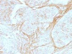
Test Specificity: This antibody recognizes a protein doublet of 20-22kDa, identified as MART-1 (Melanoma Antigen Recognized by T cells 1) or Melan-A. MART-1 is a newly identified melanocyte differentiation antigen recognized by autologous cytotoxic T lymphocytes. Seven other melanoma associated antigens recognized by autologous cytotoxic T cells include MAGE-1, MAGE-3, tyrosinase, gp100, gp75, BAGE-1, and GAGE-1. Subcellular fractionation shows that MART-1 is present in melanosomes and endoplasmic reticulum. This MAb labels melanomas and other tumors showing melanocytic differentiation. It is also a useful positive-marker for angiomyolipomas. It does not stain tumor cells of epithelial, lymphoid, glial, or mesenchymal origin.
Clone: MLANA/788
Dilution: Western Blot 0.5 - 1.0 ug/ml, Flow Cytometry 0.5 - 1 ug/million cells in 0.1 ml, Immunohistochemistry-Paraffin 0.5 - 1.0 ug/ml, Immunofluorescence 0.5 - 1.0 ug/ml
Classification: Monoclonal
Form: Purified
Regulatory Status: RUO
Target Species: Human, Mouse, Rat, Canine
Gene Accession No.: Q16655
Gene ID (Entrez): 2315
Immunogen: Recombinant human MLANA protein
Primary or Secondary: Primary
Content And Storage: Store at 4C.
Antigen: Melanoma Associated Antigen (PNL2)
Concentration: 0.2mg/mL
Applications: Immunohistochemistry (Paraffin), SDS-Page, Immunofluorescence
Conjugate: Unconjugated
Host Species: Mouse
Research Discipline: __
Formulation: 10mM PBS and 0.05% BSA with 0.05% Sodium Azide
Gene Alias: __
Gene Symbols: __
Isotype: IgG1
Purification Method: Protein A or G purified
Test Specificity: Anti-PNL2 is a novel monoclonal antibody, which has recently been introduced as an immunohistochemical reagent to stain melanocytes and tumors derived therefrom. The antigen recognized by PNL2 is different from Melan A and gp100. Its epitope is not destroyed by digestion with neuraminidase i.e. its epitope id not glycosylated. Anti-PNL2 may be most useful because of its high sensitivity for metastatic melanoma (87%), as opposed to 76% for anti-HMB45 and 82% for anti-MART-1. Anti-PNL2 labels intra-epidermal nevi while the dermal component of compound nevi are largely non-reactive with anti-PNL2. Antibodies against PNL2, MART-1 (Melan A) and HMB45 stain most clear cell sarcoma cells and a few cells in angio-myolipomas and lymphangioleiomyomatosis. Anti-PNL2 is a useful antibody for the identification of melanomas and clear cell sarcomas. Differential diagnosis is aided by the results from a panel of antibodies, including antibodies against HMB45, MART-1, tyrosinase, and MiTF.
Clone: PNL2
Dilution: Immunohistochemistry-Paraffin 0.5 - 1.0 ug/ml, SDS-Page, Immunofluorescence 0.5 - 1.0 ug/ml
Classification: Monoclonal
Form: Purified
Regulatory Status: RUO
Target Species: Human, Mouse, Canine
Gene Accession No.: __
Gene ID (Entrez): __
Immunogen: Melanocyte antigen
Primary or Secondary: Primary
Content And Storage: Store at 4C.