Melanoma Associated Antigen (PNL2) Antibody (PNL2), Novus Biologicals™
Mouse Monoclonal Antibody
Manufacturer: Fischer Scientific
The price for this product is unavailable. Please request a quote
Antigen
Melanoma Associated Antigen (PNL2)
Concentration
0.2mg/mL
Applications
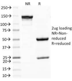
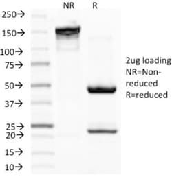
Immunohistochemistry (Paraffin), SDS-Page, Immunofluorescence
Conjugate
Unconjugated
Host Species
Mouse
Target Species
Human, Mouse, Canine
Immunogen
Melanocyte antigen
Primary or Secondary
Primary
Content And Storage
Store at 4C.
Clone
PNL2
Dilution
Immunohistochemistry-Paraffin 0.5 - 1.0 ug/ml, SDS-Page, Immunofluorescence 0.5 - 1.0 ug/ml
Classification
Monoclonal
Form
Purified
Regulatory Status
RUO
Formulation
10mM PBS and 0.05% BSA with 0.05% Sodium Azide
Isotype
IgG1
Purification Method
Protein A or G purified
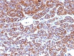
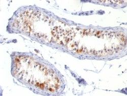
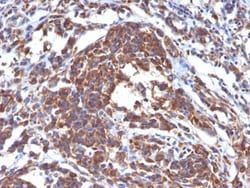
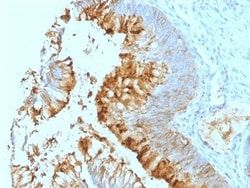
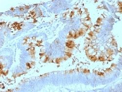
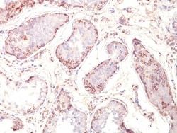
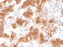
Test Specificity
Anti-PNL2 is a novel monoclonal antibody, which has recently been introduced as an immunohistochemical reagent to stain melanocytes and tumors derived therefrom. The antigen recognized by PNL2 is different from Melan A and gp100. Its epitope is not destroyed by digestion with neuraminidase i.e. its epitope id not glycosylated. Anti-PNL2 may be most useful because of its high sensitivity for metastatic melanoma (87%), as opposed to 76% for anti-HMB45 and 82% for anti-MART-1. Anti-PNL2 labels intra-epidermal nevi while the dermal component of compound nevi are largely non-reactive with anti-PNL2. Antibodies against PNL2, MART-1 (Melan A) and HMB45 stain most clear cell sarcoma cells and a few cells in angio-myolipomas and lymphangioleiomyomatosis. Anti-PNL2 is a useful antibody for the identification of melanomas and clear cell sarcomas. Differential diagnosis is aided by the results from a panel of antibodies, including antibodies against HMB45, MART-1, tyrosinase, and MiTF.
Related Products
Description
- Ensure accurate, reproducible results in Immunohistochemistry (Paraffin), Immunofluorescence Melanoma Associated Antigen (PNL2) Monoclonal specifically detects Melanoma Associated Antigen (PNL2) in Human, Canine samples
- It is validated for Immunohistochemistry, Immunocytochemistry/Immunofluorescence, Immunohistochemistry-Paraffin, Immunofluorescence.
Compare Similar Items
Show Difference
Antigen: Melanoma Associated Antigen (PNL2)
Concentration: 0.2mg/mL
Applications: Immunohistochemistry (Paraffin), SDS-Page, Immunofluorescence
Conjugate: Unconjugated
Host Species: Mouse
Target Species: Human, Mouse, Canine
Immunogen: Melanocyte antigen
Primary or Secondary: Primary
Content And Storage: Store at 4C.
Clone: PNL2
Dilution: Immunohistochemistry-Paraffin 0.5 - 1.0 ug/ml, SDS-Page, Immunofluorescence 0.5 - 1.0 ug/ml
Classification: Monoclonal
Form: Purified
Regulatory Status: RUO
Formulation: 10mM PBS and 0.05% BSA with 0.05% Sodium Azide
Isotype: IgG1
Purification Method: Protein A or G purified
Test Specificity: Anti-PNL2 is a novel monoclonal antibody, which has recently been introduced as an immunohistochemical reagent to stain melanocytes and tumors derived therefrom. The antigen recognized by PNL2 is different from Melan A and gp100. Its epitope is not destroyed by digestion with neuraminidase i.e. its epitope id not glycosylated. Anti-PNL2 may be most useful because of its high sensitivity for metastatic melanoma (87%), as opposed to 76% for anti-HMB45 and 82% for anti-MART-1. Anti-PNL2 labels intra-epidermal nevi while the dermal component of compound nevi are largely non-reactive with anti-PNL2. Antibodies against PNL2, MART-1 (Melan A) and HMB45 stain most clear cell sarcoma cells and a few cells in angio-myolipomas and lymphangioleiomyomatosis. Anti-PNL2 is a useful antibody for the identification of melanomas and clear cell sarcomas. Differential diagnosis is aided by the results from a panel of antibodies, including antibodies against HMB45, MART-1, tyrosinase, and MiTF.
Antigen: MFG-E8
Concentration: 0.2mg/mL
Applications: Flow Cytometry, Immunohistochemistry (Paraffin), Immunofluorescence
Conjugate: Unconjugated
Host Species: Mouse
Target Species: Human
Immunogen: Human milk fat globule membrane preparation
Primary or Secondary: Primary
Content And Storage: Store at 4C.
Clone: SPM291
Dilution: Flow Cytometry 0.5 - 1 ug/million cells in 0.1 ml, Immunohistochemistry-Paraffin 0.5 - 1.0 ug/ml, Immunofluorescence 1 - 2 ug/ml
Classification: Monoclonal
Form: Purified
Regulatory Status: RUO
Formulation: 10mM PBS and 0.05% BSA with 0.05% Sodium Azide
Isotype: IgG1 κ
Purification Method: Protein A or G purified
Test Specificity: Recognizes a protein of 40-45kDa, identified as human milk fat globule membrane protein (HMFG). HMFG is present on normal human breast epithelial cells and cell lines derived from breast carcinomas, as well as to the outer surface of the human milk fat globule. HMFG is considered as a differentiation marker. It is useful as specific breast epithelial marker and can also provide a tool to study the role of the cell surface in normal and neoplastic mammary development.
Antigen: MITF
Concentration: 0.2mg/mL
Applications: Flow Cytometry, Immunohistochemistry (Paraffin), SDS-Page, Immunofluorescence
Conjugate: Unconjugated
Host Species: Mouse
Target Species: Human, Mouse (Negative), Rat (Negative)
Immunogen: NH2 terminus fragment of human Mi protein
Primary or Secondary: Primary
Content And Storage: Store at 4C.
Clone: D5
Dilution: Flow Cytometry 0.5 - 1 ug/million cells in 0.1 ml, Immunohistochemistry-Paraffin 0.5 - 1.0 ug/ml, SDS-Page, Immunofluorescence 0.5 - 1.0 ug/ml
Classification: Monoclonal
Form: Purified
Regulatory Status: RUO
Formulation: 10mM PBS and 0.05% BSA with 0.05% Sodium Azide
Isotype: IgG1 κ
Purification Method: Protein A or G purified
Test Specificity: MITF (microphthalmia transcription factor) is a basic helix-loop-helix-leucine-zipper (bHLH-Zip) transcription factor that regulates the development and survival of melanocytes and retinal pigment epithelium, and also is involved in transcription of pigmentation enzyme genes such as tyrosinase TRP1 and TRP2. MITF has been shown to be phosphorylated by MAP kinase in response to c-kit activation, resulting in upregulation of MITF transcriptional activity. Mutations of the MITF gene are associated with the autosomal dominant hereditary deafness and pigmentation condition, Waardenburg Syndrome type 2A. Multiple isoforms of MITF exist, including MITF-A, MITF-B, MITF-C, MITF-H, and MITF-M, which differ in the amino-terminal domain and in their expression patterns. The MITF-M isoform is restricted to the melanocyte cell lineage. Anti-MITF, D5, recognizes a nuclear protein, which is expressed in the majority of primary and metastatic epithelioid malignant melanomas as well as in normal melanoc