TRIM29 Antibody (TRIM29/1042), Novus Biologicals™
Mouse Monoclonal Antibody
Manufacturer: Fischer Scientific
The price for this product is unavailable. Please request a quote
Antigen
TRIM29
Concentration
0.2mg/mL
Applications
Western Blot, Flow Cytometry, SDS-Page, Immunofluorescence
Conjugate
Unconjugated
Host Species
Mouse
Target Species
Human
Gene Accession No.
Q14134
Gene ID (Entrez)
23650
Immunogen
Recombinant fragment (126 Amino acid residues between aa 1-200) of human TRIM29 protein
Primary or Secondary
Primary
Content And Storage
Store at 4C.
Molecular Weight of Antigen
66 kDa
Clone
TRIM29/1042
Dilution
Western Blot 0.5 - 1.0 ug/ml, Flow Cytometry 0.5 - 1 ug/million cells in 0.1 ml, SDS-Page, Immunofluorescence 0.5 - 1.0 ug/ml
Classification
Monoclonal
Form
Purified
Regulatory Status
RUO
Formulation
1.0mM PBS and 0.05% BSA with 0.05% Sodium Azide
Gene Alias
Ataxia telangiectasia group D-associated protein, ATDCataxia-telangiectasia group D-associated protein, FLJ36085, tripartite motif containing 29, tripartite motif protein TRIM29, tripartite motif-containing 29, tripartite motif-containing protein 29
Gene Symbols
TRIM29
Isotype
IgG2b κ
Purification Method
Protein A or G purified
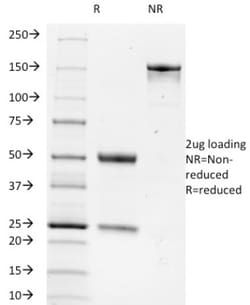
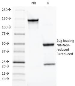
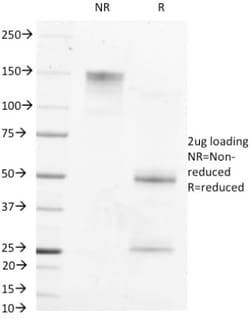
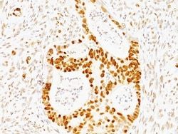
Test Specificity
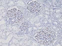
It recognizes a 66kDa protein, which is identified as Tripartite motif-containing protein 29 (TRIM29). It interacts with the intermediate filament protein vimentin, a substrate for the PKC family of protein kinases, and with hPKCI-1, an inhibitor of the PKCs. TRIM29 protein contains both zinc finger and leucine zipper motifs, suggesting that the it may form homodimers and possibly associate with DNA. High expression of TRIM29 has been reported in gastric cancer and pancreatic cancer, and correlates with enhanced tumor growth and lymph node metastasis. TRIM29 is also able to distinguish lung squamous cell carcinoma from lung adenocarcinoma with ∼90% positive accuracy, when used in a panel with TTF-1, p63, CK5/6, and Napsin-A antibodies.
Related Products
Description
- Ensure accurate, reproducible results in Western Blot, Flow Cytometry, Immunofluorescence TRIM29 Monoclonal specifically detects TRIM29 in Human samples
- It is validated for Western Blot.
Compare Similar Items
Show Difference
Antigen: TRIM29
Concentration: 0.2mg/mL
Applications: Western Blot, Flow Cytometry, SDS-Page, Immunofluorescence
Conjugate: Unconjugated
Host Species: Mouse
Target Species: Human
Gene Accession No.: Q14134
Gene ID (Entrez): 23650
Immunogen: Recombinant fragment (126 Amino acid residues between aa 1-200) of human TRIM29 protein
Primary or Secondary: Primary
Content And Storage: Store at 4C.
Molecular Weight of Antigen: 66 kDa
Clone: TRIM29/1042
Dilution: Western Blot 0.5 - 1.0 ug/ml, Flow Cytometry 0.5 - 1 ug/million cells in 0.1 ml, SDS-Page, Immunofluorescence 0.5 - 1.0 ug/ml
Classification: Monoclonal
Form: Purified
Regulatory Status: RUO
Formulation: 1.0mM PBS and 0.05% BSA with 0.05% Sodium Azide
Gene Alias: Ataxia telangiectasia group D-associated protein, ATDCataxia-telangiectasia group D-associated protein, FLJ36085, tripartite motif containing 29, tripartite motif protein TRIM29, tripartite motif-containing 29, tripartite motif-containing protein 29
Gene Symbols: TRIM29
Isotype: IgG2b κ
Purification Method: Protein A or G purified
Test Specificity: It recognizes a 66kDa protein, which is identified as Tripartite motif-containing protein 29 (TRIM29). It interacts with the intermediate filament protein vimentin, a substrate for the PKC family of protein kinases, and with hPKCI-1, an inhibitor of the PKCs. TRIM29 protein contains both zinc finger and leucine zipper motifs, suggesting that the it may form homodimers and possibly associate with DNA. High expression of TRIM29 has been reported in gastric cancer and pancreatic cancer, and correlates with enhanced tumor growth and lymph node metastasis. TRIM29 is also able to distinguish lung squamous cell carcinoma from lung adenocarcinoma with ∼90% positive accuracy, when used in a panel with TTF-1, p63, CK5/6, and Napsin-A antibodies.
Antigen: __
Concentration: __
Applications: __
Conjugate: __
Host Species: __
Target Species: __
Gene Accession No.: __
Gene ID (Entrez): __
Immunogen: __
Primary or Secondary: __
Content And Storage: __
Molecular Weight of Antigen: __
Clone: __
Dilution: __
Classification: __
Form: __
Regulatory Status: __
Formulation: __
Gene Alias: __
Gene Symbols: __
Isotype: __
Purification Method: __
Test Specificity: __

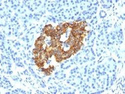
Antigen: Tyrosinase
Concentration: 0.2mg/mL
Applications: Flow Cytometry, Immunohistochemistry (Paraffin), Immunofluorescence
Conjugate: Unconjugated
Host Species: Mouse
Target Species: Human
Gene Accession No.: __
Gene ID (Entrez): 7299
Immunogen: Recombinant human TYR protein
Primary or Secondary: Primary
Content And Storage: Store at 4C.
Molecular Weight of Antigen: 75 kDa
Clone: OCA1/812
Dilution: Flow Cytometry 0.5 - 1 ug/million cells in 0.1 ml, Immunohistochemistry-Paraffin 0.5 - 1.0 ug/ml, Immunofluorescence 1 - 2 ug/ml
Classification: Monoclonal
Form: Purified
Regulatory Status: RUO
Formulation: 10mM PBS and 0.05% BSA with 0.05% Sodium Azide
Gene Alias: CMM8, EC 1.14.18.1, LB24-AB, Monophenol monooxygenase, OCA1A, OCAIA, SHEP3, SK29-AB, Tumor rejection antigen AB, tyrosinase, tyrosinase (oculocutaneous albinism IA)
Gene Symbols: TYR
Isotype: IgG2a κ
Purification Method: Protein A or G purified
Test Specificity: Recognizes a cluster of proteins between 70-80kDa, identified as tyrosinase. Occasionally a minor band at 55kDa is also detected. This MAb shows no cross-reaction with MAGE-1 and tyrosinase-related protein 1, TRP-1/gp75. Tyrosinase is a copper-containing metalloglycoprotein that catalyzes several steps in the melanin pigment biosynthetic pathway; the hydroxylation of tyrosine to L-3,4-dihydroxy-phenylalanine (dopa), and the subsequent oxidation of dopa to dopaquinone. Mutations of the tyrosinase gene occur in various forms of albinism. Tyrosinase is one of the targets for cytotoxic T-cell recognition in melanoma patients. Staining of melanomas with this MAb shows tyrosinase in melanotic as well as amelanotic variants. This MAb is a useful marker for melanocytes and melanomas.
