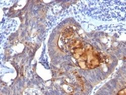
Tyrosinase Antibody (OCA1/812), Novus Biologicals™
Mouse Monoclonal Antibody
Manufacturer: Fischer Scientific
The price for this product is unavailable. Please request a quote
Antigen
Tyrosinase
Concentration
0.2mg/mL
Applications
Flow Cytometry, Immunohistochemistry (Paraffin), Immunofluorescence
Conjugate
Unconjugated
Host Species
Mouse
Research Discipline
Lipid and Metabolism
Formulation
10mM PBS and 0.05% BSA with 0.05% Sodium Azide
Gene ID (Entrez)
7299
Immunogen
Recombinant human TYR protein
Primary or Secondary
Primary
Content And Storage
Store at 4C.
Molecular Weight of Antigen
75 kDa
Clone
OCA1/812
Dilution
Flow Cytometry 0.5 - 1 ug/million cells in 0.1 ml, Immunohistochemistry-Paraffin 0.5 - 1.0 ug/ml, Immunofluorescence 1 - 2 ug/ml
Classification
Monoclonal
Form
Purified
Regulatory Status
RUO
Target Species
Human
Gene Alias
CMM8, EC 1.14.18.1, LB24-AB, Monophenol monooxygenase, OCA1A, OCAIA, SHEP3, SK29-AB, Tumor rejection antigen AB, tyrosinase, tyrosinase (oculocutaneous albinism IA)
Gene Symbols
TYR
Isotype
IgG2a κ
Purification Method
Protein A or G purified
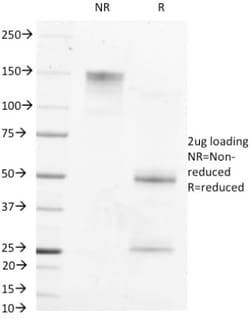
Test Specificity
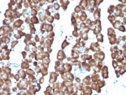
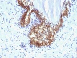
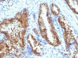
Recognizes a cluster of proteins between 70-80kDa, identified as tyrosinase. Occasionally a minor band at 55kDa is also detected. This MAb shows no cross-reaction with MAGE-1 and tyrosinase-related protein 1, TRP-1/gp75. Tyrosinase is a copper-containing metalloglycoprotein that catalyzes several steps in the melanin pigment biosynthetic pathway; the hydroxylation of tyrosine to L-3,4-dihydroxy-phenylalanine (dopa), and the subsequent oxidation of dopa to dopaquinone. Mutations of the tyrosinase gene occur in various forms of albinism. Tyrosinase is one of the targets for cytotoxic T-cell recognition in melanoma patients. Staining of melanomas with this MAb shows tyrosinase in melanotic as well as amelanotic variants. This MAb is a useful marker for melanocytes and melanomas.
Related Products


Description
- Ensure accurate, reproducible results in ELISA, Flow Cytometry, Immunohistochemistry (Paraffin), Immunofluorescence Tyrosinase Monoclonal specifically detects Tyrosinase in Human samples
- It is validated for Flow Cytometry, ELISA, Immunohistochemistry, Immunocytochemistry/Immunofluorescence, Immunohistochemistry-Paraffin, Immunofluorescence.
Compare Similar Items
Show Difference

Antigen: Tyrosinase
Concentration: 0.2mg/mL
Applications: Flow Cytometry, Immunohistochemistry (Paraffin), Immunofluorescence
Conjugate: Unconjugated
Host Species: Mouse
Research Discipline: Lipid and Metabolism
Formulation: 10mM PBS and 0.05% BSA with 0.05% Sodium Azide
Gene ID (Entrez): 7299
Immunogen: Recombinant human TYR protein
Primary or Secondary: Primary
Content And Storage: Store at 4C.
Molecular Weight of Antigen: 75 kDa
Clone: OCA1/812
Dilution: Flow Cytometry 0.5 - 1 ug/million cells in 0.1 ml, Immunohistochemistry-Paraffin 0.5 - 1.0 ug/ml, Immunofluorescence 1 - 2 ug/ml
Classification: Monoclonal
Form: Purified
Regulatory Status: RUO
Target Species: Human
Gene Alias: CMM8, EC 1.14.18.1, LB24-AB, Monophenol monooxygenase, OCA1A, OCAIA, SHEP3, SK29-AB, Tumor rejection antigen AB, tyrosinase, tyrosinase (oculocutaneous albinism IA)
Gene Symbols: TYR
Isotype: IgG2a κ
Purification Method: Protein A or G purified
Test Specificity: Recognizes a cluster of proteins between 70-80kDa, identified as tyrosinase. Occasionally a minor band at 55kDa is also detected. This MAb shows no cross-reaction with MAGE-1 and tyrosinase-related protein 1, TRP-1/gp75. Tyrosinase is a copper-containing metalloglycoprotein that catalyzes several steps in the melanin pigment biosynthetic pathway; the hydroxylation of tyrosine to L-3,4-dihydroxy-phenylalanine (dopa), and the subsequent oxidation of dopa to dopaquinone. Mutations of the tyrosinase gene occur in various forms of albinism. Tyrosinase is one of the targets for cytotoxic T-cell recognition in melanoma patients. Staining of melanomas with this MAb shows tyrosinase in melanotic as well as amelanotic variants. This MAb is a useful marker for melanocytes and melanomas.
Antigen: Tyrosinase
Concentration: 0.2mg/mL
Applications: Flow Cytometry, Immunohistochemistry (Paraffin), Immunofluorescence
Conjugate: Unconjugated
Host Species: Mouse
Research Discipline: Lipid and Metabolism
Formulation: 10mM PBS and 0.05% BSA with 0.05% Sodium Azide
Gene ID (Entrez): 7299
Immunogen: Recombinant tyrosinase protein (T311); Recombinant human TYR protein (OCA1/812)
Primary or Secondary: Primary
Content And Storage: Store at 4C.
Molecular Weight of Antigen: 75 kDa
Clone: T311 + OCA1/812
Dilution: Flow Cytometry 0.5 - 1 ug/million cells in 0.1 ml, Immunohistochemistry-Paraffin 0.5 - 1.0 ug/ml, Immunofluorescence 1 - 2 ug/ml
Classification: Monoclonal
Form: Purified
Regulatory Status: RUO
Target Species: Human
Gene Alias: CMM8, EC 1.14.18.1, LB24-AB, Monophenol monooxygenase, OCA1A, OCAIA, SHEP3, SK29-AB, Tumor rejection antigen AB, tyrosinase, tyrosinase (oculocutaneous albinism IA)
Gene Symbols: TYR
Isotype: IgG
Purification Method: Protein A or G purified
Test Specificity: Recognizes a cluster of proteins between 70-80kDa, identified as tyrosinase. Occasionally a minor band at 55kDa is also detected. This MAb shows no cross-reaction with MAGE-1 and tyrosinase-related protein 1, TRP-1/gp75. Tyrosinase is a copper-containing metalloglycoprotein that catalyzes several steps in the melanin pigment biosynthetic pathway; the hydroxylation of tyrosine to L-3,4-dihydroxy-phenylalanine (dopa), and the subsequent oxidation of dopa to dopaquinone. Mutations of the tyrosinase gene occur in various forms of albinism. Tyrosinase is one of the targets for cytotoxic T-cell recognition in melanoma patients. Staining of melanomas with this MAb shows tyrosinase in melanotic as well as amelanotic variants. This MAb is a useful marker for melanocytes and melanomas.
Antigen: ECM-1/Secretory Component P85
Concentration: 0.2mg/mL
Applications: Flow Cytometry, Immunohistochemistry (Paraffin), SDS-Page, Immunofluorescence
Conjugate: Unconjugated
Host Species: Mouse
Research Discipline: Extracellular Matrix, Immunology
Formulation: 10mM PBS and 0.05% BSA with 0.05% Sodium Azide
Gene ID (Entrez): 1893
Immunogen: Recombinant human ECM1 protein
Primary or Secondary: Primary
Content And Storage: Store at 4C.
Molecular Weight of Antigen: __
Clone: ECM1/792
Dilution: Flow Cytometry 0.5 - 1 ug/million cells in 0.1 ml, Immunohistochemistry-Paraffin 0.5 - 1.0 ug/ml, SDS-Page, Immunofluorescence 1 - 2 ug/ml
Classification: Monoclonal
Form: Purified
Regulatory Status: RUO
Target Species: Human, Rat
Gene Alias: ECM1, extracellular matrix protein 1, Secretory Component Glycoprotein, Secretory component p85
Gene Symbols: ECM1
Isotype: IgG1 κ
Purification Method: Protein A or G purified
Test Specificity: This MAb reacts with a reduction-resistant epitope present in both free and SIgA bound Secretory Component. It does not react with the cell lines lacking secretory component. The antibody is useful for studying the distribution and level of both free and bound secretory component. Secretory component is differentially expressed in epithelium, and the antibody is a popular marker for identifying subpopulations of epithelial cells and epithelial differentiation. The Secretory component antibody is a useful research tool for studying mucosal immunity, inflammation, remodeling, differentiation and tumorigenesis, all processes associated with differential secretory component expression.