Ornithine Decarboxylase Antibody (ODC1/487), Novus Biologicals™
Mouse Monoclonal Antibody
Manufacturer: Fischer Scientific
The price for this product is unavailable. Please request a quote
Antigen
Ornithine Decarboxylase
Concentration
0.2mg/mL
Applications
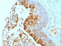
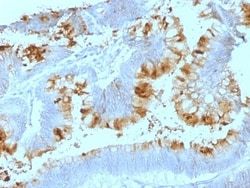
Western Blot, Flow Cytometry, Immunohistochemistry (Paraffin), SDS-Page, Immunofluorescence
Conjugate
Unconjugated
Host Species
Mouse
Research Discipline
Cell Cycle and Replication
Formulation
10mM PBS and 0.05% BSA with 0.05% Sodium Azide
Gene Alias
ODCEC 4.1.1.17, ornithine decarboxylase, ornithine decarboxylase 1
Gene Symbols
ODC1
Isotype
IgG2a κ
Purification Method
Protein A or G purified
Test Specificity
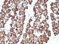
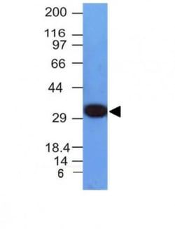
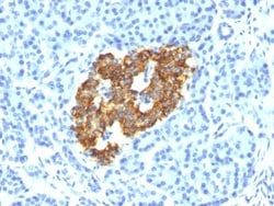
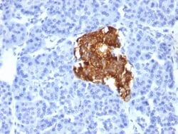
Recognizes a 53kDa protein, identified as the Ornithine Decarboxylase (ODC-1). ODC is the initial and rate-limiting enzyme in the biosynthetic pathway of polyamines and is involved in the conversion of ornithine to putrescine. The biological activity of ODC-1 is rapidly induced in response to virtually all agents known to promote cell proliferation including hormones, drugs, growth factors, mitogens, and tumor promoters. Reportedly, ODC mRNA levels are elevated in lung carcinomas as well as in colon adenomas and carcinomas. ODC activity in colorectal carcinomas is greater than those in adenomas and normal mucosa.
Clone
ODC1/487
Dilution
Western Blot 0.5 - 1.0 ug/ml, Flow Cytometry 0.5 - 1 ug/million cells in 0.1 ml, Immunohistochemistry-Paraffin 0.5 - 1.0 ug/ml, SDS-Page, Immunofluorescence 0.5 - 1.0 ug/ml
Classification
Monoclonal
Form
Purified
Regulatory Status
RUO
Target Species
Human, Rat
Gene Accession No.
P11926
Gene ID (Entrez)
4953
Immunogen
Recombinant human ODC-1 protein
Primary or Secondary
Primary
Content And Storage
Store at 4C.
Molecular Weight of Antigen
53 kDa
Related Products
Description
- Ensure accurate, reproducible results in Western Blot, Flow Cytometry, Immunohistochemistry (Paraffin), Immunofluorescence Ornithine Decarboxylase Monoclonal specifically detects Ornithine Decarboxylase in Human, Mouse, Rat samples
- It is validated for Western Blot, Flow Cytometry, Immunohistochemistry, Immunocytochemistry/Immunofluorescence, Immunohistochemistry-Paraffin.
Compare Similar Items
Show Difference
Antigen: Ornithine Decarboxylase
Concentration: 0.2mg/mL
Applications: Western Blot, Flow Cytometry, Immunohistochemistry (Paraffin), SDS-Page, Immunofluorescence
Conjugate: Unconjugated
Host Species: Mouse
Research Discipline: Cell Cycle and Replication
Formulation: 10mM PBS and 0.05% BSA with 0.05% Sodium Azide
Gene Alias: ODCEC 4.1.1.17, ornithine decarboxylase, ornithine decarboxylase 1
Gene Symbols: ODC1
Isotype: IgG2a κ
Purification Method: Protein A or G purified
Test Specificity: Recognizes a 53kDa protein, identified as the Ornithine Decarboxylase (ODC-1). ODC is the initial and rate-limiting enzyme in the biosynthetic pathway of polyamines and is involved in the conversion of ornithine to putrescine. The biological activity of ODC-1 is rapidly induced in response to virtually all agents known to promote cell proliferation including hormones, drugs, growth factors, mitogens, and tumor promoters. Reportedly, ODC mRNA levels are elevated in lung carcinomas as well as in colon adenomas and carcinomas. ODC activity in colorectal carcinomas is greater than those in adenomas and normal mucosa.
Clone: ODC1/487
Dilution: Western Blot 0.5 - 1.0 ug/ml, Flow Cytometry 0.5 - 1 ug/million cells in 0.1 ml, Immunohistochemistry-Paraffin 0.5 - 1.0 ug/ml, SDS-Page, Immunofluorescence 0.5 - 1.0 ug/ml
Classification: Monoclonal
Form: Purified
Regulatory Status: RUO
Target Species: Human, Rat
Gene Accession No.: P11926
Gene ID (Entrez): 4953
Immunogen: Recombinant human ODC-1 protein
Primary or Secondary: Primary
Content And Storage: Store at 4C.
Molecular Weight of Antigen: 53 kDa

Antigen: VCAM-1/CD106
Concentration: 0.2 mg/mL
Applications: Flow Cytometry, SDS-Page, Immunofluorescence
Conjugate: Unconjugated
Host Species: Mouse
Research Discipline: Cancer, Mesenchymal Stem Cell Markers, Stem Cell Markers
Formulation: 10mM PBS and 0.05% BSA with 0.05% Sodium Azide
Gene Alias: CD106, CD106 antigen, DKFZp779G2333, INCAM-100, L1CAM, MGC99561, vascular cell adhesion molecule 1, vascular cell adhesion protein 1, V-CAM 1, VCAM1, VCAM-1
Gene Symbols: VCAM1
Isotype: IgG1 κ
Purification Method: Protein A or G purified
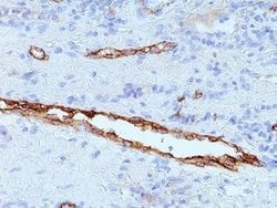
Test Specificity: Recognizes a protein of 110kDa, identified as CD106 (also known as vascular cell adhesion molecule-1 (VCAM-1) and INCAM-100). CD106 is a member of the Ig superfamily of adhesion molecules and is expressed at high levels on cytokine stimulated vascular endothelial cells, and at minimal levels on un-stimulated endothelial cells. It is also present on follicular and inter-follicular dendritic cells of lymph nodes, myoblasts, and some macrophages. CD106 serves as a ligand for leukocyte integrin (VLA-4 or CD49d/CD29) and mediates cell adhesion of leukocytes to activated endothelium. It plays a role in various immunological and inflammatory responses. This MAb inhibits the binding of leukocytes to VCAM-1 on stimulated endothelial cells.
Clone: B-K9
Dilution: Flow Cytometry 0.5 - 1 ug/million cells in 0.1 ml, SDS-Page, Immunofluorescence 1 - 2 ug/ml
Classification: Monoclonal
Form: Purified
Regulatory Status: RUO
Target Species: Human
Gene Accession No.: P19320
Gene ID (Entrez): 7412
Immunogen: Activated human umbilical vein endothelial cells (HUVEC)
Primary or Secondary: Primary
Content And Storage: Store at 4C.
Molecular Weight of Antigen: 110 kDa
Antigen: Vimentin
Concentration: 0.2mg/mL
Applications: Flow Cytometry, Immunohistochemistry (Paraffin), Immunofluorescence
Conjugate: Unconjugated
Host Species: Mouse
Research Discipline: Cancer, Cellular Markers, Cytoskeleton Markers, Growth and Development, Hypoxia, Neuronal Cell Markers, Neuronal Stem Cell Markers, Neuroscience, Signal Transduction, Stem Cell Markers, Stem Cells
Formulation: 10mM PBS and 0.05% BSA with 0.05% Sodium Azide
Gene Alias: FLJ36605, vimentin
Gene Symbols: VIM
Isotype: IgM
Purification Method: Protein A or G purified
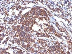
Test Specificity: This MAb reacts with a 58kDa protein identified as vimentin. It reacts with a non-hematopoietic epitope of vimentin and shows no cross-reaction with other closely related intermediate filament proteins (IFPs) such as desmin, keratin, neurofilament, and glial fibrillary acid protein.Vimentin is ubiquitously expressed in mesenchymal cells such as fibroblasts, smooth muscle cells, and endothelium. Antibody against vimentin is useful as part of an antibody panel for differential diagnosis of tumors of unknown origin.Ab-2 does not react with leukocyte common antigen-positive tissues such as lymphomas and leukemias.
Clone: LN-6
Dilution: Flow Cytometry 0.5 - 1.0 ug/million cells in 0.1 ml, Immunohistochemistry-Paraffin 1 - 2 ug/ml, Immunofluorescence 0.5 - 1.0 ug/ml
Classification: Monoclonal
Form: Purified
Regulatory Status: RUO
Target Species: Human, Mouse, Rat, Porcine, Feline, Primate, Rabbit, Sheep
Gene Accession No.: P08670
Gene ID (Entrez): 7431
Immunogen: Human thymic nuclear extract
Primary or Secondary: Primary
Content And Storage: Store at 4C.
Molecular Weight of Antigen: __