NGFR/TNFRSF16/p75NTR Antibody (NGFR5 + NTR/912), Novus Biologicals™
Mouse Monoclonal Antibody
Manufacturer: Fischer Scientific
The price for this product is unavailable. Please request a quote
Antigen
NGF R/TNFRSF16/p75NTR
Concentration
0.2mg/mL
Applications
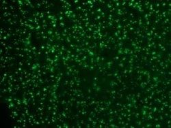
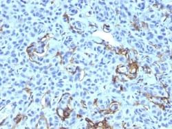
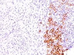
Flow Cytometry, Immunohistochemistry (Paraffin), Immunofluorescence
Conjugate
Unconjugated
Host Species
Mouse
Research Discipline
Apoptosis, Neuronal Cell Markers, Neuronal Stem Cell Markers, Neuroscience, Stem Cell Markers
Formulation
10mM PBS and 0.05% BSA with 0.05% Sodium Azide
Gene Alias
CD271, CD271 antigen, Gp80-LNGFR, member 16, nerve growth factor receptor, NGF receptor, p75 ICD, TNFRSF16nerve growth factor receptor (TNFR superfamily, member 16), tumor necrosis factor receptor superfamily member 16
Gene Symbols
NGFR
Isotype
IgG
Purification Method
Protein A or G purified
Test Specificity
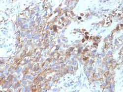
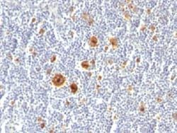
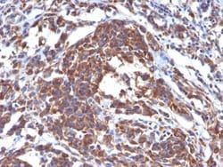
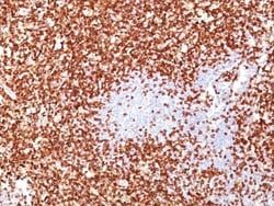
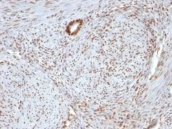
It recognizes a glycoprotein of 75kDa, identified as low affinity Nerve Growth Factor (NGF) Receptor (p75NGFR) or Neurotrophin Receptor (p75NTR). NGFR is expressed in various neural crest cells and their tumors such as melanocytes, melanomas, neuroblastomas, pheochromocytomas and neurofibromas. Reportedly, anti-NGFR is a reliable marker for desmoplastic and neurotropic melanomas. NGFR is expressed in mature non-neural cells such as perivascular cells, dental pulp cells, lymphoidal follicular dendritic cells, basal epithelium of oral mucosa and hair follicles, prostate basal cells, and myoepithelial cells. Anti-NGFR stains the myoepithelial cells of breast ducts and intra-lobular fibroblasts of breast ducts.
Clone
NGFR5 + NTR/912
Dilution
Flow Cytometry 0.5 - 1 ug/million cells in 0.1 ml, Immunohistochemistry-Paraffin 0.5 - 1.0 ug/ml, Immunofluorescence 1 - 2 ug/ml
Classification
Monoclonal
Form
Purified
Regulatory Status
RUO
Target Species
Human, Primate, Mouse (Negative), Rat (Negative)
Gene Accession No.
P08138
Gene ID (Entrez)
4804
Immunogen
NGFR from A875 melanoma cells (NGFR5); Recombinant human p75 NGFR protein (NTR/912)
Primary or Secondary
Primary
Content And Storage
Store at 4C.
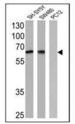
Molecular Weight of Antigen
75 kDa
Related Products

Description
- Ensure accurate, reproducible results in Flow Cytometry, Immunohistochemistry (Paraffin), Immunofluorescence NGFR/TNFRSF16/p75NTR Monoclonal specifically detects NGFR/TNFRSF16/p75NTR in Human, Primate, Mouse (Negative), Rat (Negative) samples
- It is validated for Flow Cytometry, Immunohistochemistry, Immunocytochemistry/Immunofluorescence, Immunohistochemistry-Paraffin, Immunofluorescence.
Compare Similar Items
Show Difference
Antigen: NGF R/TNFRSF16/p75NTR
Concentration: 0.2mg/mL
Applications: Flow Cytometry, Immunohistochemistry (Paraffin), Immunofluorescence
Conjugate: Unconjugated
Host Species: Mouse
Research Discipline: Apoptosis, Neuronal Cell Markers, Neuronal Stem Cell Markers, Neuroscience, Stem Cell Markers
Formulation: 10mM PBS and 0.05% BSA with 0.05% Sodium Azide
Gene Alias: CD271, CD271 antigen, Gp80-LNGFR, member 16, nerve growth factor receptor, NGF receptor, p75 ICD, TNFRSF16nerve growth factor receptor (TNFR superfamily, member 16), tumor necrosis factor receptor superfamily member 16
Gene Symbols: NGFR
Isotype: IgG
Purification Method: Protein A or G purified
Test Specificity: It recognizes a glycoprotein of 75kDa, identified as low affinity Nerve Growth Factor (NGF) Receptor (p75NGFR) or Neurotrophin Receptor (p75NTR). NGFR is expressed in various neural crest cells and their tumors such as melanocytes, melanomas, neuroblastomas, pheochromocytomas and neurofibromas. Reportedly, anti-NGFR is a reliable marker for desmoplastic and neurotropic melanomas. NGFR is expressed in mature non-neural cells such as perivascular cells, dental pulp cells, lymphoidal follicular dendritic cells, basal epithelium of oral mucosa and hair follicles, prostate basal cells, and myoepithelial cells. Anti-NGFR stains the myoepithelial cells of breast ducts and intra-lobular fibroblasts of breast ducts.
Clone: NGFR5 + NTR/912
Dilution: Flow Cytometry 0.5 - 1 ug/million cells in 0.1 ml, Immunohistochemistry-Paraffin 0.5 - 1.0 ug/ml, Immunofluorescence 1 - 2 ug/ml
Classification: Monoclonal
Form: Purified
Regulatory Status: RUO
Target Species: Human, Primate, Mouse (Negative), Rat (Negative)
Gene Accession No.: P08138
Gene ID (Entrez): 4804
Immunogen: NGFR from A875 melanoma cells (NGFR5); Recombinant human p75 NGFR protein (NTR/912)
Primary or Secondary: Primary
Content And Storage: Store at 4C.
Molecular Weight of Antigen: 75 kDa
Antigen: NGF R/TNFRSF16/p75NTR
Concentration: 0.2mg/mL
Applications: Flow Cytometry, Immunocytochemistry, Immunofluorescence, Immunohistochemistry (Paraffin)
Conjugate: Unconjugated
Host Species: Mouse
Research Discipline: Apoptosis, Neuronal Cell Markers, Neuronal Stem Cell Markers, Neuroscience, Stem Cell Markers
Formulation: 10mM PBS and 0.05% BSA with 0.05% Sodium Azide
Gene Alias: CD271, CD271 antigen, Gp80-LNGFR, member 16, nerve growth factor receptor, NGF receptor, p75 ICD, TNFRSF16nerve growth factor receptor (TNFR superfamily, member 16), tumor necrosis factor receptor superfamily member 16
Gene Symbols: NGFR
Isotype: IgG1 κ
Purification Method: Protein A or G purified
Test Specificity: It recognizes a glycoprotein of 75kDa, identified as low affinity Nerve Growth Factor (NGF) Receptor (p75NGFR) or Neurotrophin Receptor (p75NTR). Its epitope spans in aa 1-160 of extracellular domain of NGFR/NTR. NGF-receptor contains an extracellular domain containing four 40-amino acid repeats with 6 cysteine residues at conserved positions followed by a serine/threonine-rich region, a single transmembrane domain, and a 155-amino acid cytoplasmic domain. The cysteine-rich region contains the nerve growth factor binding domain. NGF is important for the development, differentiation, and survival of variety of neuronal and non-neuronal cells. Its action is mediated by binding two distinct receptors, the high affinity p140 and low affinity p75.
Clone: NGFR5
Dilution: Flow Cytometry 0.5 - 1 ug/million cells in 0.1 ml, Immunocytochemistry/Immunofluorescence 1:10-1:500, Immunohistochemistry-Paraffin 0.5 - 1.0 ug/ml, Immunofluorescence 1 - 2 ug/ml
Classification: Monoclonal
Form: Purified
Regulatory Status: RUO
Target Species: Human, Baboon, Feline, Ferret, Monkey, Rabbit, Mouse (Negative), Rat (Negative)
Gene Accession No.: P08138
Gene ID (Entrez): 4804
Immunogen: NGFR from A875 melanoma cells
Primary or Secondary: Primary
Content And Storage: Store at 4C.
Molecular Weight of Antigen: 75 kDa
Antigen: Nucleolin
Concentration: 0.2mg/mL
Applications: Western Blot, Flow Cytometry, Immunohistochemistry (Paraffin), Immunofluorescence
Conjugate: Unconjugated
Host Species: Mouse
Research Discipline: Cell Cycle and Replication, Chromatin Research, Core ESC Like Genes, DNA Repair, DNA replication Transcription Translation and Splicing, Stem Cell Markers
Formulation: 10mM PBS and 0.05% BSA with 0.05% Sodium Azide
Gene Alias: C23, FLJ45706, FLJ59041, nucleolin, Protein C23
Gene Symbols: NCL
Isotype: IgG
Purification Method: Protein A or G purified
Test Specificity: Recognizes a protein of ∼76kDa, which is identified as Nucleolin (NCL). It is the major nucleolar phosphoprotein of growing eukaryotic cells. NCL is located mainly in dense fibrillar regions of the nucleolus. It is found associated with intranucleolar chromatin and pre-ribosomal particles. Human NCL gene consists of 14 exons with 13 introns and spans approximately 11kb. It induces chromatin decondensation by binding to histone H1. It is thought to play a role in pre-rRNA transcription and ribosome assembly. This MAb can be used to stain the nucleoli in cell or tissue preparations and can be used as a marker of the nucleoli in subcellular fractions. It produces a speckled pattern in the nuclei of cells of normal and malignant cells and may be used to stain the nucleoli of cells in fixed or frozen tissue sections. It can be used with paraformaldehyde fixed frozen tissue or cell preparations and formalin fixed, paraffin-embedded tissue sections.
Clone: 364-5 + NCL/902
Dilution: Western Blot 0.5 - 1.0 ug/ml, Flow Cytometry 0.5 - 1 ug/million cells in 0.1 ml, Immunohistochemistry-Paraffin 0.5 - 1.0 ug/ml, Immunofluorescence 0.5 - 1.0 ug/ml
Classification: Monoclonal
Form: Purified
Regulatory Status: RUO
Target Species: Human, Bovine (Negative), Mouse (Negative), Rat (Negative)
Gene Accession No.: P19338
Gene ID (Entrez): 4691
Immunogen: Lysate of SU-DHL-1 Nuclei (364-5); Recombinant human NCL protein (NCL/902)
Primary or Secondary: Primary
Content And Storage: Store at 4C.
Molecular Weight of Antigen: 76 kDa