NCAM-1/CD56 Antibody (123C3.D5 + 123A8), Novus Biologicals™
Mouse Monoclonal Antibody
Manufacturer: Fischer Scientific
The price for this product is unavailable. Please request a quote
Antigen
NCAM-1/CD56
Dilution
Western Blot 0.5-1ug/ml, Flow Cytometry 0.5-1ug/million cells, Immunocytochemistry/Immunofluorescence 1-2ug/ml, Immunoprecipitation 1-2ug/500ug protein lysate, Immunohistochemistry-Paraffin 0.5-1ug/ml, Immunohistochemistry-Frozen 0.5-1ug/mlimmunohistochemistry-Paraffin 0.5-1ug/ml
Classification
Monoclonal
Form
Purified
Regulatory Status
RUO
Target Species
Human
Gene Accession No.
P13591
Gene ID (Entrez)
4684
Immunogen
Membrane preparation of a small cell lung carcinoma
Primary or Secondary
Primary
Content And Storage
Store at 4C.
Clone
123c3.D5+123A8
Applications
Western Blot, Flow Cytometry, Immunocytochemistry, Immunofluorescence, Immunoprecipitation, Immunohistochemistry (Paraffin)
Conjugate
Unconjugated
Host Species
Mouse
Research Discipline
Astrocyte Markers, Cellular Markers, Cytokine Research, Cytoskeleton Markers, Growth and Development, Hematopoietic Stem Cell Markers, Immunology, Innate Immunity, Membrane Vesicle Markers, Neuronal Cell Markers, Neuronal Stem Cell Markers, Neuroscience, Stem Cell Markers
Formulation
10mM PBS and 0.05% BSA with 0.05% Sodium Azide
Gene Alias
CD56, CD56/ NCAM-1, CD56 antigen, MSK39, N-CAM-1, NCAM-1, NCAMantigen recognized by monoclonal 5.1H11, neural cell adhesion molecule 1, neural cell adhesion molecule, NCAM
Gene Symbols
NCAM1
Isotype
IgG
Purification Method
Protein A purified
Test Specificity
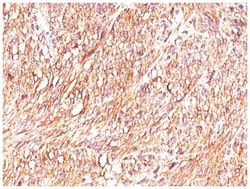
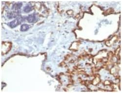
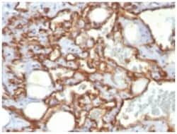
This MAb reacts with an extracellular domain (close to transmembrane) of CD56/NCAM. Three isoforms of neural cell adhesion molecule (NCAM) are produced by differential splicing of the RNA transcript from a single gene. The 135kDa isoform is the basic molecule, which is glycosylated or sialylated to produce the mature species. Anti-CD56 recognizes two proteins of the neural cell adhesion molecule, the basic molecule expressed on most neuroectodermally derived tissues and neoplasms (e.g. retinoblastoma, medulloblastomas, astrocytomas, neuroblastomas, and small cell carcinomas). It is also expressed on some mesodermally derived tumors (rhabdomyosarcoma). Anti-CD56 plays an important role in the diagnosis of nodal and nasal NK/T-cell lymphomas.
Related Products

Description
- NCAM-1/CD56 Monoclonal specifically detects NCAM-1/CD56 in Human samples
- It is validated for Flow Cytometry, Immunohistochemistry, Immunocytochemistry/Immunofluorescence, Immunohistochemistry-Paraffin.
Compare Similar Items
Show Difference
Antigen: NCAM-1/CD56
Dilution: Western Blot 0.5-1ug/ml, Flow Cytometry 0.5-1ug/million cells, Immunocytochemistry/Immunofluorescence 1-2ug/ml, Immunoprecipitation 1-2ug/500ug protein lysate, Immunohistochemistry-Paraffin 0.5-1ug/ml, Immunohistochemistry-Frozen 0.5-1ug/mlimmunohistochemistry-Paraffin 0.5-1ug/ml
Classification: Monoclonal
Form: Purified
Regulatory Status: RUO
Target Species: Human
Gene Accession No.: P13591
Gene ID (Entrez): 4684
Immunogen: Membrane preparation of a small cell lung carcinoma
Primary or Secondary: Primary
Content And Storage: Store at 4C.
Clone: 123c3.D5+123A8
Applications: Western Blot, Flow Cytometry, Immunocytochemistry, Immunofluorescence, Immunoprecipitation, Immunohistochemistry (Paraffin)
Conjugate: Unconjugated
Host Species: Mouse
Research Discipline: Astrocyte Markers, Cellular Markers, Cytokine Research, Cytoskeleton Markers, Growth and Development, Hematopoietic Stem Cell Markers, Immunology, Innate Immunity, Membrane Vesicle Markers, Neuronal Cell Markers, Neuronal Stem Cell Markers, Neuroscience, Stem Cell Markers
Formulation: 10mM PBS and 0.05% BSA with 0.05% Sodium Azide
Gene Alias: CD56, CD56/ NCAM-1, CD56 antigen, MSK39, N-CAM-1, NCAM-1, NCAMantigen recognized by monoclonal 5.1H11, neural cell adhesion molecule 1, neural cell adhesion molecule, NCAM
Gene Symbols: NCAM1
Isotype: IgG
Purification Method: Protein A purified
Test Specificity: This MAb reacts with an extracellular domain (close to transmembrane) of CD56/NCAM. Three isoforms of neural cell adhesion molecule (NCAM) are produced by differential splicing of the RNA transcript from a single gene. The 135kDa isoform is the basic molecule, which is glycosylated or sialylated to produce the mature species. Anti-CD56 recognizes two proteins of the neural cell adhesion molecule, the basic molecule expressed on most neuroectodermally derived tissues and neoplasms (e.g. retinoblastoma, medulloblastomas, astrocytomas, neuroblastomas, and small cell carcinomas). It is also expressed on some mesodermally derived tumors (rhabdomyosarcoma). Anti-CD56 plays an important role in the diagnosis of nodal and nasal NK/T-cell lymphomas.
Antigen: CD31/PECAM-1
Dilution: Flow Cytometry 0.5-1ug/million cells, Immunohistochemistry, Immunocytochemistry/Immunofluorescence 0.5-1ug/ml, Immunohistochemistry-Paraffin 0.5-1ug/ml
Classification: Monoclonal
Form: Purified
Regulatory Status: RUO
Target Species: Human, Primate, Rabbit
Gene Accession No.: P16284
Gene ID (Entrez): 5175
Immunogen: Human recombinant CD31 protein (C31.3) & Membrane preparation of a spleen from a patient with hairy cell leukemia (JC/70A)
Primary or Secondary: Primary
Content And Storage: Store at 4C.
Clone: C31.3 + JC/70A
Applications: Flow Cytometry, Immunohistochemistry, Immunocytochemistry, Immunofluorescence, Immunohistochemistry (Paraffin)
Conjugate: Unconjugated
Host Species: Mouse
Research Discipline: Angiogenesis, Cancer, Cellular Markers, Cytoskeleton Markers, Embryonic Stem Cell Markers, Endothelial Cell Markers, Extracellular Matrix, Hematopoietic Stem Cell Markers, Immunology, Mesenchymal Stem Cell Markers, Myeloid Cell Markers, Signal Transduction, Stem Cell Markers
Formulation: 10mM PBS and 0.05% BSA with 0.05% Sodium Azide
Gene Alias: adhesion molecule, CD31, CD31 antigen, CD31/EndoCAM, EndoCAM, FLJ34100, FLJ58394, GPIIA', PECA1, PECAM-1, PECAM-1, CD31/EndoCAM, platelet endothelial cell adhesion molecule, platelet/endothelial cell adhesion molecule
Gene Symbols: PECAM1
Isotype: IgG
Purification Method: Protein A or G purified
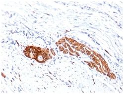
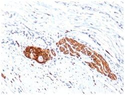
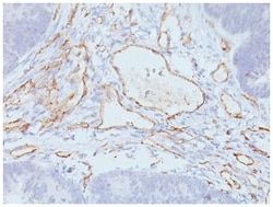
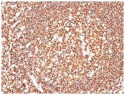
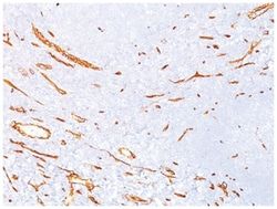
Test Specificity: CD31 (PECAM-1) is a transmembrane glycoprotein member of the immunoglobulin supergene family of adhesion molecules. CD31 is expressed by stem cells of the hematopoietic system and is primarily used to identify and concentrate these cells for experimental studies as well as for bone marrow transplantation. Anti-CD31 has shown to be highly specific and sensitive for vascular endothelial cells. Staining of nonvascular tumors (excluding hematopoietic neoplasms) is rare. CD31 MAb reacts with normal, benign, and malignant endothelial cells which make up blood vessel lining. The level of CD31 expression can help to determine the degree of tumor angiogenesis, and a high level of CD31 expression may imply a rapidly growing tumor and potentially a predictor of tumor recurrence.

Antigen: ACTH
Dilution: Western Blot 0.5-1.0ug/ml, Flow Cytometry 0.5-1ug/million cells, Immunocytochemistry/Immunofluorescence 0.5-1ug/ml, Immunoprecipitation 0.5-1ug/500ug protein lysate, Immunohistochemistry-Paraffin 0.5-1ug/ml, Immunohistochemistry-Frozen 0.5-1ug/ml
Classification: Monoclonal
Form: Purified
Regulatory Status: RUO
Target Species: Human, Mouse, Rat
Gene Accession No.: P01189
Gene ID (Entrez): 5443
Immunogen: Synthetic peptide corresponding to aa1-24 of human ACTH (AH26); N-terminal fragment of human ACTH conjugated to KLH (57)
Primary or Secondary: Primary
Content And Storage: Store at 4C.
Clone: AH26 + 57
Applications: Western Blot, Flow Cytometry, Immunocytochemistry, Immunofluorescence, Immunoprecipitation, Immunohistochemistry (Paraffin)
Conjugate: Unconjugated
Host Species: Mouse
Research Discipline: Neuroscience, Nutrient Sensing in the Brain
Formulation: PBS with 0.05% BSA. with 0.05% Sodium Azide
Gene Alias: ACTH, adrenocorticotropic hormone, adrenocorticotropin, alpha-melanocyte-stimulating hormone, alpha-MSH, beta-endorphin, beta-LPH, beta-melanocyte-stimulating hormone, beta-MSH, CLIP, corticotropin-like intermediary peptide, corticotropin-lipotropin, gamma-LPH, gamma-MSH, lipotropin beta, lipotropin gamma, LPH, melanotropin alpha, melanotropin beta, melanotropin gamma, met-enkephalin, MSH, NPP, POC, pro-ACTH-endorphin, proopiomelanocortin, pro-opiomelanocortin, proopiomelanocortin preproprotein
Gene Symbols: POMC
Isotype: IgG
Purification Method: Protein A purified
Test Specificity: ACTH (same as Corticotropin) is a 39 amino acid active peptide produced by the anterior pituitary. This MAb is specific to Synacthen (aa1-24 of ACTH); does not react with CLIP (aa17-39 of ACTH). POMC (pro-opiomelanocortin or corticotropin-lipotropin) is a 267 amino acid polypeptide hormone precursor that goes through extensive, tissue-specific posttranslational processing by convertases. POMC is cleaved into ten hormone chains named NPP, ACTH, alpha-MSH (Melanocyte Stimulating Hormone), beta-MSH, gamma-MSH, CLIP (corticotropin-like intermediary peptide), Lipotropin-beta, Lipotropin-gamma, beta-endorphin and Met-enkephalin. ACTH is also produced by cells of immune system (T-cells, B-cells, and macrophages) in response to stimuli associated with stress. Anti-ACTH is a useful marker in classification of pituitary tumors and the study of pituitary disease. It reacts with ACTH-producing cells (corticotrophs). It also may react with other tumors (e.g. some small cell carcinomas of the lung) causing paraneoplastic syndromes by secreting ACTH.