TMPRSS2, Mouse anti-Human, Clone: P5H9-A3, Millipore Sigma™
Mouse Monoclonal Antibody
Manufacturer: Fischer Scientific
The price for this product is unavailable. Please request a quote
Antigen
TMPRSS2
Dilution
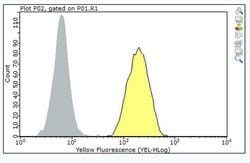
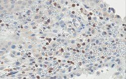
Immunohistochemistry (Paraffin) Analysis: 1:50 dilution from a representative lot detected TMPRSS2 in human kidney tissue sections.Enzyme Immunoassay (ELISA) Analysis: A representative lot detected TMPRSS2 in ELISA applications (Lucas, J.M., et. al. (2008). J Pathol. 215(2):118-25).Western Blotting Analysis: A representative lot detected TMPRSS2 in Western Blotting applications (Lucas, J.M., et. al. (2008). J Pathol. 215(2):118-25).Immunohistochemistry (Paraffin) Analysis: A representative lot detected TMPRSS2 in Immunohistochemistry applications (Lucas, J.M., et. al. (2008). J Pathol. 215(2):118-25; Bertram, S., et. al. (2012). PLoS One. 7(4):e35876).
Classification
Monoclonal
Form
Purified
Regulatory Status
RUO
Target Species
Human
Gene Alias
Transmembrane protease serine 2;Serine protease 10
Immunogen
KLH-conjugated linear peptide corresponding to 16 amino acids from the extracellular domain of human transmembrane protease serine 2.
Primary or Secondary
Primary
Content And Storage
Stable for 1 year at 2-8°C from date of receipt.
Clone
P5H9-A3
Applications
ELISA, Immunohistochemistry (Paraffin), Western Blot
Conjugate
Unconjugated
Host Species
Mouse
Research Discipline
Inflammation & Immunology
Formulation
Purified mouse monoclonal antibody IgG1 in buffer containing 0.1 M Tris-Glycine (pH 7.4), 150 mM NaCl with 0.05% sodium azide.
Gene Symbols
TMPRSS2;PRSS10
Isotype
IgG1 κ
Purification Method
Protein G purified
Test Specificity
Clone P5H9-A3 specifically detects human Transmembrane protease serine 2. It targets an epitope with in the extracellular serine protease domain.
Description
- Anti-TMPRSS2, clone P5H9-A3, Cat
- No
- MABF2158, is a mouse monoclonal antibody that detects Transmembrane protease serine 2 and has been tested for use in ELISA, Immunohistochemistry (Paraffin), and Western Blotting
- Transmembrane protease serine 2 (UniProt: O15393; also known as Serine protease 10, TMPRSS2) is encoded by the TMPRSS2 (also known as PRSS10) gene (Gene ID: 7113) in human
- TMPRSS2 is a single-pass type II membrane protein of the peptidase S1 family that is shown to proteolytically cleave and activate the viral spike glycoproteins, which facilitate virus-cell membrane fusions
- It is shown to facilitate human SARS coronavirus (SARS-CoV) infection via two independent mechanisms, proteolytic cleavage of ACE2 that promotes viral uptake and cleavage of coronavirus spike glycoprotein, which activates the glycoprotein for cathepsin L-independent host cell entry
- TMPRSS2 is highly expressed in the prostate tissue and lower expression levels are observed in the epithelia of the colon, stomach, epididymis and breast tissue
- Some expression has also been reported in pancreatic acini, hepatic bile ducts, testicular Leydig cells and the kidney
- Its expression levels are significantly elevated in both neoplastic prostate and in the epithelium of prostatic hyperplasia
- TMPRSS2 has a cytoplasmic domain (aa 1-84), a transmembrane domain (aa 85-105), and an extracellular domain (aa 106-492)
- Its peptidase S1 domain is localized to amino acids 256-489
- It is reported to be proteolytically processed by an autocatalytic mechanism generating the transmembrane protease serine 2 non-catalytic chain (1-255) and the transmembrane protease serine 2 catalytic chain (256-492)
- Two isoforms of TMPRSS2 have been described that are produced by alternative splicing.
Compare Similar Items
Show Difference

Antigen: TMPRSS2
Dilution: Immunohistochemistry (Paraffin) Analysis: 1:50 dilution from a representative lot detected TMPRSS2 in human kidney tissue sections.Enzyme Immunoassay (ELISA) Analysis: A representative lot detected TMPRSS2 in ELISA applications (Lucas, J.M., et. al. (2008). J Pathol. 215(2):118-25).Western Blotting Analysis: A representative lot detected TMPRSS2 in Western Blotting applications (Lucas, J.M., et. al. (2008). J Pathol. 215(2):118-25).Immunohistochemistry (Paraffin) Analysis: A representative lot detected TMPRSS2 in Immunohistochemistry applications (Lucas, J.M., et. al. (2008). J Pathol. 215(2):118-25; Bertram, S., et. al. (2012). PLoS One. 7(4):e35876).
Classification: Monoclonal
Form: Purified
Regulatory Status: RUO
Target Species: Human
Gene Alias: Transmembrane protease serine 2;Serine protease 10
Immunogen: KLH-conjugated linear peptide corresponding to 16 amino acids from the extracellular domain of human transmembrane protease serine 2.
Primary or Secondary: Primary
Content And Storage: Stable for 1 year at 2-8°C from date of receipt.
Clone: P5H9-A3
Applications: ELISA, Immunohistochemistry (Paraffin), Western Blot
Conjugate: Unconjugated
Host Species: Mouse
Research Discipline: Inflammation & Immunology
Formulation: Purified mouse monoclonal antibody IgG1 in buffer containing 0.1 M Tris-Glycine (pH 7.4), 150 mM NaCl with 0.05% sodium azide.
Gene Symbols: TMPRSS2;PRSS10
Isotype: IgG1 κ
Purification Method: Protein G purified
Test Specificity: Clone P5H9-A3 specifically detects human Transmembrane protease serine 2. It targets an epitope with in the extracellular serine protease domain.

Antigen: CD63 (LAMP3)
Dilution: Immunohistochemistry (Paraffin) Analysis: A 1:250 dilution from a representative lot detected CD63 (LAMP3) in human spleen and human bone marrow tissue sections.Immunocytochemistry Analysis: A representative lot detected CD63 (LAMP3) in Immunocytochemistry applications (Atkinson, B., et. al. (1984). Cancer Res. 44(6):2577-81).Flow Cytometry Analysis: A representative lot detected CD63 (LAMP3) in Flow Cytometry applications (Li, J., et. al. (2003). J Immunol. 171(6):2922-9).Western Blotting Analysis: A representative lot detected CD63 (LAMP3) in Western Blotting applications (Smith, M., et. al. (1997). Melanoma Res. 7 Suppl 2:S163-70).Immunohistochemistry Analysis: A representative lot detected CD63 (LAMP3) in Immunohistochemistry applications (Li, J., et. al. (2003). J Immunol. 171(6):2922-9).
Classification: Monoclonal
Form: Purified
Regulatory Status: RUO
Target Species: Human
Gene Alias: CD63 antigen;Granulophysin;Lysosomal-associated membrane protein 3;LAMP-3;Melanoma-associated antigen ME491;OMA81H;Ocular melanoma-associated antigen;Tetraspanin-30;Tspan-30
Immunogen: Clear supernatant from SK-Mel-23 cell lysate.
Primary or Secondary: Primary
Content And Storage: Stable for 1 year at 2-8°C from date of receipt.
Clone: ME491
Applications: Flow Cytometry, Immunocytochemistry, Immunohistochemistry (Paraffin), Western Blot
Conjugate: Unconjugated
Host Species: Mouse
Research Discipline: Inflammation & Immunology
Formulation: Purified mouse monoclonal antibody IgG1 in buffer containing 0.1 M Tris-Glycine (pH 7.4), 150 mM NaCl with 0.05% sodium azide.
Gene Symbols: CD63;MLA1;TSPAN30
Isotype: IgG1 κ
Purification Method: Protein G purified
Test Specificity: Clone ME491 specifically detects CD63 (LAMP-3) in human cells.
Antigen: HLA-F
Dilution: ELISA Analysis: A representative lot detected HLA-F in ELISA applications (Lee, N., et. al. (2010). Eur J Immunol. 40(8):2308-18).Western Blotting Analysis: A representative lot detected HLA-F in Western Blotting applications (Lee, N., et. al. (2010). Eur J Immunol. 40(8):2308-18; Ishitani, A., et. al. (2003). J Immunol. 171(3):1376-84).Flow Cytometry Analysis: A representative lot detected HLA-F in Flow Cytometry applications (Lee, N., et. al. (2010). Eur J Immunol. 40(8):2308-18).Immunohistochemistry Analysis: A representative lot detected HLA-F in Immunohistochemistry applications (Ishitani, A., et. al. (2003). J Immunol. 171(3):1376-84).
Classification: Monoclonal
Form: Purified
Regulatory Status: RUO
Target Species: Human
Gene Alias: HLA class I histocompatibility antigen alpha chain F;CDA12;HLA F antigen;Leukocyte antigen F;MHC class I antigen F
Immunogen: Full-length human recombinant HLA-F Heavy chains (excluding the signal sequence).
Primary or Secondary: Primary
Content And Storage: Stable for 1 year at 2-8°C from date of receipt.
Clone: 3D11
Applications: ELISA, Flow Cytometry, Immunohistochemistry (Paraffin), Western Blot
Conjugate: Unconjugated
Host Species: Mouse
Research Discipline: Inflammation & Immunology
Formulation: Purified mouse monoclonal antibody IgG1 in buffer containing 0.1 M Tris-Glycine (pH 7.4), 150 mM NaCl with 0.05% sodium azide.
Gene Symbols: HLA-F;HLA-5.4;HLAF
Isotype: IgG1 κ
Purification Method: Protein G purified
Test Specificity: Clone 3D11 specifically detects human HLA-F. It targets an epitope with in the alpha 2 domain from the N-terminal half.