EphA2, Mouse anti-Human, Clone: F2-27, Millipore Sigma™
Mouse Monoclonal Antibody
Manufacturer: Fischer Scientific
The price for this product is unavailable. Please request a quote
Antigen
EphA2
Dilution
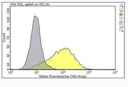
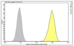
Inhibition Analysis: A representative lot inhibited cancer cell invasion in a three-dimensional spheroid formation assay. (Tanaka, T., et. al. (2017). Transl Oncol. 10(4):476-484).Immunoprecipitation Analysis: A representative lot detected EphA2 in Immunoprecipitation applications (Tanaka, T., et. al. (2017). Transl Oncol. 10(4):476-484).Flow Cytometry Analysis: A representative lot detected EphA2 in Flow Cytometry applications (Tanaka, T., et. al. (2017). Transl Oncol. 10(4):476-484).
Classification
Monoclonal
Form
Purified
Regulatory Status
RUO
Target Species
Human
Gene Alias
Ephrin type-A receptor 2;EC: 2.7.10.1;Epithelial cell kinase;Tyrosine-protein kinase receptor ECK
Gene Symbols
EPHA2;ECK
Isotype
IgG1 κ
Purification Method
Protein G purified
Test Specificity
Clone F2-27 specifically detects Ephrin type -A receptor 2 in human.
Clone
F2-27
Applications
Flow Cytometry, Immunoprecipitation, Inhibition Assays
Conjugate
Unconjugated
Host Species
Mouse
Research Discipline
Signaling
Formulation
Purified mouse monoclonal antibody IgG1 in buffer containing 0.1 M Tris-Glycine (pH 7.4), 150 mM NaCl with 0.05% sodium azide.
Gene ID (Entrez)
NP_004422
Immunogen
KP-2, KP-3, SUIT-2, and MIAPaCa-2 cells.
Primary or Secondary
Primary
Content And Storage
Stable for 1 year at 2-8°C from date of receipt.
Description
- Anti-EphA2, clone F2-27, Cat
- No
- MABS2026, is a mouse monoclonal antibody that detects Ephrin type-A receptor 2 and has been tested for use in Flow Cytometry, Immunoprecipitation, and Inhibition Assay
- Ephrin type-A receptor 2 (UniProt: P29317; also known as EC: 2.7.10.1, Epithelial cell kinase, Tyrosine-protein kinase receptor ECK) is encoded by the EPHA2 (also known as ECK) gene (Gene ID: 1969) in human
- Ephrin type-A receptor 2 is a homodimeric single-pass type I membrane protein that is expressed in brain and glioma tissue and in glioma cell lines
- It is also highly expressed in tissues that contain a high proportion of epithelial cells
- Its levels are shown to be up-regulated by UV irradiation via a TP53-independent, MAPK-dependent mechanism
- Ephrin type-A receptor 2 serves as a receptor tyrosine kinase that binds promiscuously membrane-bound ephrin-A family ligands residing on adjacent cells, leading to contact-dependent bidirectional signaling into neighboring cells
- It is involved in the regulation of migration, integrin-mediated adhesion, and in proliferation and differentiation of cells
- Ephrin type-A receptor 2 is phosphorylated on tyrosine residues upon binding and activation by EFNA1
- Phosphorylation on Tyr 588 and Tyr 594 is reported to be essential for binding VAV2 and VAV3 while phosphorylation on Tyr 735 and Tyr 930 are required for binding PI3-kinase p85 subunit
- It is synthesized with a signal peptide (aa 1-23) that is cleaved off in the mature form
- The mature form has an extracellular domain (aa 24-537), a short transmembrane domain (aa 538-558), and a cytoplasmic domain (aa 559-976)
- Two isoforms of Ephrin type-A receptor 2 have been described that are produced by alternative splicing
- EphA2 is reported to be overexpressed in skin tumors murine models and its overexpression may represent a compensatory feedback mechanism during tumorigenesis
- (Ref.: Guo, H., et al
- (2006)
- Cancer Res
- 66(14); 7050-7058).
Compare Similar Items
Show Difference
Antigen: EphA2
Dilution: Inhibition Analysis: A representative lot inhibited cancer cell invasion in a three-dimensional spheroid formation assay. (Tanaka, T., et. al. (2017). Transl Oncol. 10(4):476-484).Immunoprecipitation Analysis: A representative lot detected EphA2 in Immunoprecipitation applications (Tanaka, T., et. al. (2017). Transl Oncol. 10(4):476-484).Flow Cytometry Analysis: A representative lot detected EphA2 in Flow Cytometry applications (Tanaka, T., et. al. (2017). Transl Oncol. 10(4):476-484).
Classification: Monoclonal
Form: Purified
Regulatory Status: RUO
Target Species: Human
Gene Alias: Ephrin type-A receptor 2;EC: 2.7.10.1;Epithelial cell kinase;Tyrosine-protein kinase receptor ECK
Gene Symbols: EPHA2;ECK
Isotype: IgG1 κ
Purification Method: Protein G purified
Test Specificity: Clone F2-27 specifically detects Ephrin type -A receptor 2 in human.
Clone: F2-27
Applications: Flow Cytometry, Immunoprecipitation, Inhibition Assays
Conjugate: Unconjugated
Host Species: Mouse
Research Discipline: Signaling
Formulation: Purified mouse monoclonal antibody IgG1 in buffer containing 0.1 M Tris-Glycine (pH 7.4), 150 mM NaCl with 0.05% sodium azide.
Gene ID (Entrez): NP_004422
Immunogen: KP-2, KP-3, SUIT-2, and MIAPaCa-2 cells.
Primary or Secondary: Primary
Content And Storage: Stable for 1 year at 2-8°C from date of receipt.

Antigen: SREBP-1
Dilution: Western Blotting Analysis: A representative lot detected SREBP-1 in Western Blotting applications (Toth, J.I., et. al. (2004). Mol Cell Biol. 24(18):8288-300; Key, C.C., et. al. (2017). Cell Rep. 19(10):2116-2129; Rosenfeld, J.M., et. al. (1998). J Biol Chem. 273(26):16112-21; Sato, R., et. al. (1994). J Biol Chem. 269(25):17267-73).
Classification: Monoclonal
Form: Purified
Regulatory Status: RUO
Target Species: Hamster, Canine, Human
Gene Alias: Sterol regulatory element-binding protein 1;Class D basic helix-loop-helix protein 1;bHLHd1;Sterol regulatory element-binding transcription factor 1
Gene Symbols: SREBF1;BHLHD1;SREBP1
Isotype: IgG1 κ
Purification Method: Protein G purified
Test Specificity: Clone 2A4 is a mouse monoclonal antibody that detects Sterol regulatory element-binding protein 1. It targets an epitope within 107 amino acids from the N-terminal half.
Clone: 2A4
Applications: Western Blot
Conjugate: Unconjugated
Host Species: Mouse
Research Discipline: Signaling
Formulation: Purified mouse monoclonal antibody IgG1 in buffer containing 0.1 M Tris-Glycine (pH 7.4), 150 mM NaCl with 0.05% sodium azide.
Gene ID (Entrez): NP_001005291.1
Immunogen: His-tagged recombinant fragment corresponding to 107 amino acids from the N-terminal half of human Sterol regulatory element-binding protein 1 (SREBP-1).
Primary or Secondary: Primary
Content And Storage: Stable for 1 year at 2-8°C from date of receipt.

Antigen: GCP2
Dilution: Western Blotting Analysis: 1μg/mL from a representative lot detected GCP2 in human glioblastoma T98G cell lysate.Immunoprecipitation Analysis: A representative lot immunoprecipitated GCP2 in Immunoprecipitation applications (Draberova, E., et. al. (2015). J Neuropathol Exp Neurol. 74(7):723-42).Immunocytochemistry Analysis: A representative lot detected GCP2 in Immunocytochemistry applications (Draberova, E., et. al. (2015). J Neuropathol Exp Neurol. 74(7):723-42).Western Blotting Analysis: A representative lot detected GCP2 in Western Blotting applications (Draberova, E., et. al. (2015). J Neuropathol Exp Neurol. 74(7):723-42).ELISA Analysis: A representative lot detected GCP2 in ELISA applications (Draberova, E., et. al. (2015). J Neuropathol Exp Neurol. 74(7):723-42).Electron Microscopy Analysis: A representative lot detected GCP2 in Electron Microscopy applications (Draberova, E., et. al. (2015). J Neuropathol Exp Neurol. 74(7):723-42). Immunoprecipitation Analysis: A represneta
Classification: Monoclonal
Form: Purified
Regulatory Status: RUO
Target Species: Mouse, Chicken, Human, Frog, Fish
Gene Alias: Gamma-tubulin complex component 2
Gene Symbols: Tubgcp2;Gcp2
Isotype: IgG1 κ
Purification Method: Protein G purified
Test Specificity: Clone GCP2-02 specifically detects GCP2 in T98G glioblastoma cells. It targets an epitope within 12 amino acids from the N-terminal region.
Clone: GCP2-02
Applications: ELISA, Immunocytochemistry, Immunomicroscopy, Immunoprecipitation, Western Blot
Conjugate: Unconjugated
Host Species: Mouse
Research Discipline: Cell Structure
Formulation: Purified mouse monoclonal antibody IgG1 in buffer containing 0.1 M Tris-Glycine (pH 7.4), 150 mM NaCl with 0.05% sodium azide.
Gene ID (Entrez): NP_001272936.1
Immunogen: GST-tagged recombinant fragment corresponding to the first 194 amino acids from the N-terminal region of murine Gamma-tubulin complex component 2.
Primary or Secondary: Primary
Content And Storage: Stable for 1 year at 2-8°C from date of receipt.