Carbonic Anhydrase IX/CA9 Antibody (66.4.C2 (PN-15)), Novus Biologicals™
Manufacturer: Fischer Scientific
Select a Size
| Pack Size | SKU | Availability | Price |
|---|---|---|---|
| Each of 1 | NBP21519902-Each-of-1 | In Stock | ₹ 56,159.00 |
NBP21519902 - Each of 1
In Stock
Quantity
1
Base Price: ₹ 56,159.00
GST (18%): ₹ 10,108.62
Total Price: ₹ 66,267.62
Antigen
Renal Cell Carcinoma (gp200)
Classification
Monoclonal
Concentration
0.2mg/mL
Dilution
Western Blot 0.5-1ug/ml, Flow Cytometry 0.5-1ug/million cells, ELISA 1-5ug/ml for coating, Immunocytochemistry/Immunofluorescence 1-2ug/ml, Immunoprecipitation 1-2ug/500ug protein lysate, Immunohistochemistry-Paraffin 0.5-1ug/ml, Immunohistochemistry-Frozen 0.5-1ug/ml
Gene Accession No.
Q16790
Gene Symbols
LY75
Immunogen
Fresh, normal human renal cortical tissue homogenate was used as immunogen to generate the RCC antibody.
Quantity
0.2 mg
Research Discipline
Dendritic Cell Markers, Ovarian Carcinoma Cell Markers, Renal Cell Carcinoma Cell Markers
Gene ID (Entrez)
4065
Target Species
Human, Equine
Form
Purified
Applications
Western Blot, Flow Cytometry, ELISA, Immunocytochemistry, Immunofluorescence, Immunoprecipitation, Immunohistochemistry (Paraffin)
Clone
66.4.C2 (PN-15)
Conjugate
Unconjugated
Formulation
PBS with 0.05% BSA. with 0.05% Sodium Azide
Gene Alias
CD205LY-75, CLEC13BLy-75, C-type lectin domain family 13 member B, DEC-205CD205 antigen, GP200-MR6, lymphocyte antigen 75
Host Species
Mouse
Purification Method
Protein G purified
Regulatory Status
RUO
Primary or Secondary
Primary
Test Specificity
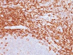
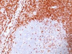
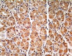
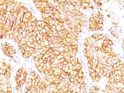
Recognizes a glycoprotein of ∼200kDa, identified as carbonic anhydrase IX (CAIX/gp200). Its epitope resides in the carbohydrate domain of gp200. It shows no significant cross-reactivity with other carbohydrate determinants, such as the Lewis blood group antigens, epithelial membrane antigen, HMFG, and AB blood group antigens. In normal kidney, gp200 is localized along the brush border of the pars convoluta and pars recta segments of the proximal tubule, as well as focally along the luminal surface of Bowman's capsule adjoining the outgoing proximal tubule. Reportedly, gp200 is expressed by 93% of primary and 84% of metastatic renal cell carcinomas. This MAb may be useful in the investigations of carcinomas of proximal nephrogenic differentiation especially those showing tubular differentiation.
Content And Storage
Store at 4C.
Isotype
IgG2b κ
Related Products


Description
- Carbonic Anhydrase IX/CA9 Monoclonal specifically detects Carbonic Anhydrase IX/CA9 in Human, Equine samples
- It is validated for Western Blot, Flow Cytometry, Immunohistochemistry, Immunocytochemistry/Immunofluorescence, Immunohistochemistry-Paraffin.
Compare Similar Items
Show Difference
Antigen: Renal Cell Carcinoma (gp200)
Classification: Monoclonal
Concentration: 0.2mg/mL
Dilution: Western Blot 0.5-1ug/ml, Flow Cytometry 0.5-1ug/million cells, ELISA 1-5ug/ml for coating, Immunocytochemistry/Immunofluorescence 1-2ug/ml, Immunoprecipitation 1-2ug/500ug protein lysate, Immunohistochemistry-Paraffin 0.5-1ug/ml, Immunohistochemistry-Frozen 0.5-1ug/ml
Gene Accession No.: Q16790
Gene Symbols: LY75
Immunogen: Fresh, normal human renal cortical tissue homogenate was used as immunogen to generate the RCC antibody.
Quantity: 0.2 mg
Research Discipline: Dendritic Cell Markers, Ovarian Carcinoma Cell Markers, Renal Cell Carcinoma Cell Markers
Gene ID (Entrez): 4065
Target Species: Human, Equine
Form: Purified
Applications: Western Blot, Flow Cytometry, ELISA, Immunocytochemistry, Immunofluorescence, Immunoprecipitation, Immunohistochemistry (Paraffin)
Clone: 66.4.C2 (PN-15)
Conjugate: Unconjugated
Formulation: PBS with 0.05% BSA. with 0.05% Sodium Azide
Gene Alias: CD205LY-75, CLEC13BLy-75, C-type lectin domain family 13 member B, DEC-205CD205 antigen, GP200-MR6, lymphocyte antigen 75
Host Species: Mouse
Purification Method: Protein G purified
Regulatory Status: RUO
Primary or Secondary: Primary
Test Specificity: Recognizes a glycoprotein of ∼200kDa, identified as carbonic anhydrase IX (CAIX/gp200). Its epitope resides in the carbohydrate domain of gp200. It shows no significant cross-reactivity with other carbohydrate determinants, such as the Lewis blood group antigens, epithelial membrane antigen, HMFG, and AB blood group antigens. In normal kidney, gp200 is localized along the brush border of the pars convoluta and pars recta segments of the proximal tubule, as well as focally along the luminal surface of Bowman's capsule adjoining the outgoing proximal tubule. Reportedly, gp200 is expressed by 93% of primary and 84% of metastatic renal cell carcinomas. This MAb may be useful in the investigations of carcinomas of proximal nephrogenic differentiation especially those showing tubular differentiation.
Content And Storage: Store at 4C.
Isotype: IgG2b κ
Antigen: Bcl-2
Classification: Monoclonal
Concentration: 0.2mg/mL
Dilution: Western Blot 0.5-1ug/ml, Flow Cytometry 0.5-1ug/million cells, ELISA 1-5ug/ml for coating, Immunocytochemistry/Immunofluorescence 1-2ug/ml, Immunoprecipitation 1-2ug/500ug protein lysate, Immunohistochemistry-Paraffin 0.5-1ug/ml, Immunohistochemistry-Frozen 0.5-1ug/ml
Gene Accession No.: P10415
Gene Symbols: BCL2
Immunogen: The synthetic peptide corresponding to amino acids range from 41-64 (GAAPAPGIFSSQPG-Cys), of human Bcl-2 was used as immunogen to generate the antibody; GenBank no. NP_000648.2 (Pezzella et al, 1990).
Quantity: 0.1 mg
Research Discipline: Apoptosis, Autophagy, Cancer, Cellular Markers, Mitochondrial Mediated Pathway, Mitophagy, Tumor Suppressors
Gene ID (Entrez): 596
Target Species: Human, Mouse (Negative), Rat (Negative)
Form: Purified
Applications: Western Blot, Flow Cytometry, ELISA, Immunocytochemistry, Immunofluorescence, Immunoprecipitation, Immunohistochemistry (Paraffin)
Clone: 100/D5
Conjugate: Unconjugated
Formulation: PBS with 0.05% BSA. with 0.05% Sodium Azide
Gene Alias: apoptosis regulator Bcl-2, B-cell CLL/lymphoma 2, Bcl-2
Host Species: Mouse
Purification Method: Protein G purified
Regulatory Status: RUO
Primary or Secondary: Primary
Test Specificity: This antibody recognizes a protein of 25-26kDa, identified as the bcl-2alpha oncoprotein. It shows no cross-reaction with Bcl-x or Bax protein. Expression of bcl-2alpha oncoprotein inhibits the programmed cell death (apoptosis). In most follicular lymphomas, neoplastic germinal centers express high levels of bcl-2alpha protein, whereas the normal or hyperplastic germinal centers are negative. Consequently, this antibody is valuable when distinguishing between reactive and neoplastic follicular proliferation in lymph node biopsies. It may also be used in distinguishing between those follicular lymphomas that express bcl-2 protein and the small number in which the neoplastic cells are bcl-2 negative.
Content And Storage: Store at 4C.
Isotype: IgG1 κ
Antigen: Bcl-2
Classification: Monoclonal
Concentration: 0.2mg/mL
Dilution: Western Blot 0.5-1ug/ml, Flow Cytometry 0.5-1ug/million cells, ELISA 1-5ug/ml for coating, Immunocytochemistry/Immunofluorescence 1-2ug/ml, Immunoprecipitation 1-2ug/500ug protein lysate, Immunohistochemistry-Paraffin 0.5-1ug/ml, Immunohistochemistry-Frozen 0.5-1ug/ml
Gene Accession No.: P10415
Gene Symbols: BCL2
Immunogen: The synthetic peptide corresponding to amino acids range from 41-64 (GAAPAPGIFSSQPG-Cys), of human Bcl-2 was used as immunogen to generate the antibody; GenBank no. NP_000648.2 (Pezzella et al, 1990).
Quantity: 0.2 mg
Research Discipline: Apoptosis, Autophagy, Cancer, Cellular Markers, Mitochondrial Mediated Pathway, Mitophagy, Tumor Suppressors
Gene ID (Entrez): 596
Target Species: Human, Mouse (Negative), Rat (Negative)
Form: Purified
Applications: Western Blot, Flow Cytometry, ELISA, Immunocytochemistry, Immunofluorescence, Immunoprecipitation, Immunohistochemistry (Paraffin)
Clone: 100/D5
Conjugate: Unconjugated
Formulation: PBS with 0.05% BSA. with 0.05% Sodium Azide
Gene Alias: apoptosis regulator Bcl-2, B-cell CLL/lymphoma 2, Bcl-2
Host Species: Mouse
Purification Method: Protein G purified
Regulatory Status: RUO
Primary or Secondary: Primary
Test Specificity: This antibody recognizes a protein of 25-26kDa, identified as the bcl-2alpha oncoprotein. It shows no cross-reaction with Bcl-x or Bax protein. Expression of bcl-2alpha oncoprotein inhibits the programmed cell death (apoptosis). In most follicular lymphomas, neoplastic germinal centers express high levels of bcl-2alpha protein, whereas the normal or hyperplastic germinal centers are negative. Consequently, this antibody is valuable when distinguishing between reactive and neoplastic follicular proliferation in lymph node biopsies. It may also be used in distinguishing between those follicular lymphomas that express bcl-2 protein and the small number in which the neoplastic cells are bcl-2 negative.
Content And Storage: Store at 4C.
Isotype: IgG1 κ