Cytokeratin 19 Antibody (A53-B/A2.26), Novus Biologicals™
Manufacturer: Fischer Scientific
Select a Size
| Pack Size | SKU | Availability | Price |
|---|---|---|---|
| Each of 1 | NBP21518602-Each-of-1 | In Stock | ₹ 54,379.00 |
NBP21518602 - Each of 1
In Stock
Quantity
1
Base Price: ₹ 54,379.00
GST (18%): ₹ 9,788.22
Total Price: ₹ 64,167.22
Antigen
Cytokeratin 19
Classification
Monoclonal
Concentration
0.2mg/mL
Dilution
Western Blot 0.5-1ug/ml, Simple Western 10 ug/ml, Flow Cytometry 0.5-1ug/million cells, ELISA 1-5ug/ml for coating, Immunocytochemistry/Immunofluorescence 1-2ug/ml, Immunoprecipitation 1-2ug/500ug protein lysate, Immunohistochemistry-Paraffin 0.5-1ug/ml, Immunohistochemistry-Frozen 0.5-1ug/ml
Gene Accession No.
P08727
Gene Symbols
KRT19
Immunogen
Human breast cancer MCF-7 cells were used as the immunogen for the Keratin-19 antibody.
Quantity
0.2 mg
Research Discipline
Cancer, Cell Biology, Cellular Markers, Cytoskeleton Markers, Neuroscience, Stem Cell Markers
Gene ID (Entrez)
3880
Target Species
Human
Form
Purified
Applications
Western Blot, Flow Cytometry, ELISA, Immunocytochemistry, Immunofluorescence, Immunoprecipitation
Clone
A53-B/A2.26
Conjugate
Unconjugated
Formulation
PBS with 0.05% BSA. with 0.05% Sodium Azide
Gene Alias
CK19, CK-19, cytokeratin 19, Cytokeratin-19,40-kDa keratin intermediate filament, K19cytokeratin-19, K1CS, keratin 19, keratin, type I cytoskeletal 19, keratin, type I, 40-kd, keratin-19, MGC15366
Host Species
Mouse
Purification Method
Protein G purified
Regulatory Status
RUO
Primary or Secondary
Primary
Test Specificity
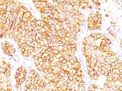
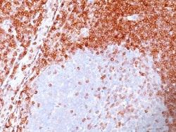
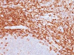
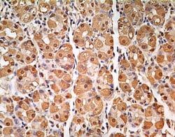
This MAb reacts with the rod domain of human cytokeratin-19 (CK19), a polypeptide of 40kDa. Its epitope maps between amino acid 312-335. CK19 is expressed in sweat gland, mammary gland ductal and secretory cells, bile ducts, gastrointestinal tract, bladder urothelium, oral epithelia, esophagus, and ectocervical epithelium. Anti-CK19 reacts with a wide variety of epithelial malignancies including adenocarcinomas of the colon, stomach, pancreas, biliary tract, liver, and breast. Perhaps the most useful application is the identification of thyroid carcinoma of the papillary type, although 50%-60% of follicular carcinomas are also labeled. Anti-CK19 is a useful marker for detection of tumor cells in lymph nodes, peripheral blood, bone marrow and breast cancer.
Content And Storage
Store at 4C.
Isotype
IgG2a κ
Related Products

Description
- Cytokeratin 19 Monoclonal specifically detects Cytokeratin 19 in Human samples
- It is validated for Western Blot, Simple Western, Flow Cytometry, Immunohistochemistry, Immunocytochemistry/Immunofluorescence, Immunohistochemistry-Paraffin, Immunohistochemistry-Frozen.
Compare Similar Items
Show Difference

Antigen: Cytokeratin 19
Classification: Monoclonal
Concentration: 0.2mg/mL
Dilution: Western Blot 0.5-1ug/ml, Simple Western 10 ug/ml, Flow Cytometry 0.5-1ug/million cells, ELISA 1-5ug/ml for coating, Immunocytochemistry/Immunofluorescence 1-2ug/ml, Immunoprecipitation 1-2ug/500ug protein lysate, Immunohistochemistry-Paraffin 0.5-1ug/ml, Immunohistochemistry-Frozen 0.5-1ug/ml
Gene Accession No.: P08727
Gene Symbols: KRT19
Immunogen: Human breast cancer MCF-7 cells were used as the immunogen for the Keratin-19 antibody.
Quantity: 0.2 mg
Research Discipline: Cancer, Cell Biology, Cellular Markers, Cytoskeleton Markers, Neuroscience, Stem Cell Markers
Gene ID (Entrez): 3880
Target Species: Human
Form: Purified
Applications: Western Blot, Flow Cytometry, ELISA, Immunocytochemistry, Immunofluorescence, Immunoprecipitation
Clone: A53-B/A2.26
Conjugate: Unconjugated
Formulation: PBS with 0.05% BSA. with 0.05% Sodium Azide
Gene Alias: CK19, CK-19, cytokeratin 19, Cytokeratin-19,40-kDa keratin intermediate filament, K19cytokeratin-19, K1CS, keratin 19, keratin, type I cytoskeletal 19, keratin, type I, 40-kd, keratin-19, MGC15366
Host Species: Mouse
Purification Method: Protein G purified
Regulatory Status: RUO
Primary or Secondary: Primary
Test Specificity: This MAb reacts with the rod domain of human cytokeratin-19 (CK19), a polypeptide of 40kDa. Its epitope maps between amino acid 312-335. CK19 is expressed in sweat gland, mammary gland ductal and secretory cells, bile ducts, gastrointestinal tract, bladder urothelium, oral epithelia, esophagus, and ectocervical epithelium. Anti-CK19 reacts with a wide variety of epithelial malignancies including adenocarcinomas of the colon, stomach, pancreas, biliary tract, liver, and breast. Perhaps the most useful application is the identification of thyroid carcinoma of the papillary type, although 50%-60% of follicular carcinomas are also labeled. Anti-CK19 is a useful marker for detection of tumor cells in lymph nodes, peripheral blood, bone marrow and breast cancer.
Content And Storage: Store at 4C.
Isotype: IgG2a κ
Antigen: CD43/Sialophorin
Classification: Monoclonal
Concentration: 0.2mg/mL
Dilution: Western Blot 0.5-1ug/ml, Flow Cytometry 0.5-1ug/million cells, ELISA 1-5ug/ml for coating, Immunocytochemistry/Immunofluorescence 1-2ug/ml, Immunoprecipitation 1-2ug/500ug protein lysate, Immunohistochemistry-Paraffin 0.5-1ug/ml, Immunohistochemistry-Frozen 0.5-1ug/ml
Gene Accession No.: P16150
Gene Symbols: SPN
Immunogen: Myeloblastic KG1 cells were used as the immunogen for this antibody.
Quantity: 0.1 mg
Research Discipline: B Cell Development and Differentiation Markers, Immunology
Gene ID (Entrez): 6693
Target Species: Human
Form: Purified
Applications: Western Blot, Flow Cytometry, ELISA, Immunocytochemistry, Immunofluorescence, Immunoprecipitation, Immunohistochemistry (Paraffin)
Clone: DF-T1
Conjugate: Unconjugated
Formulation: PBS with 0.05% BSA. with 0.05% Sodium Azide
Gene Alias: CD43 antigen, CD43), Galactoglycoprotein, GALGP, Leukocyte sialoglycoprotein, Sialophorin, sialophorin (gpL115, leukosialin, CD43)
Host Species: Mouse
Purification Method: Protein G purified
Regulatory Status: RUO
Primary or Secondary: Primary
Test Specificity: It recognizes a cell surface glycoprotein of 95/115/135kDa (depending upon the extent of glycosylation), identified as CD43. 70-90% of T-cell lymphomas and from 22-37% of B-cell lymphomas express CD43. No reactivity has been observed with reactive B-cells. So a B-lineage population that co-expresses CD43 is highly likely to be a malignant lymphoma, especially a low-grade lymphoma, rather than a reactive B-cell population. When CD43 antibody is used in combination with anti-CD20, effective immunophenotyping of the lymphomas in formalin-fixed tissues can be obtained. Co-staining of a lymphoid infiltrate with anti-CD20 and anti-CD43 argues against a reactive process and favors a diagnosis of lymphoma.
Content And Storage: Store at 4C.
Isotype: IgG1 κ
Antigen: CD43/Sialophorin
Classification: Monoclonal
Concentration: 0.2mg/mL
Dilution: Western Blot 0.5-1ug/ml, Flow Cytometry 0.5-1ug/million cells, ELISA 1-5ug/ml for coating, Immunocytochemistry/Immunofluorescence 1-2ug/ml, Immunoprecipitation 1-2ug/500ug protein lysate, Immunohistochemistry-Paraffin 0.5-1ug/ml, Immunohistochemistry-Frozen 0.5-1ug/ml
Gene Accession No.: P16150
Gene Symbols: SPN
Immunogen: Myeloblastic KG1 cells were used as the immunogen for this antibody.
Quantity: 0.2 mg
Research Discipline: B Cell Development and Differentiation Markers, Immunology
Gene ID (Entrez): 6693
Target Species: Human
Form: Purified
Applications: Western Blot, Flow Cytometry, ELISA, Immunocytochemistry, Immunofluorescence, Immunoprecipitation, Immunohistochemistry (Paraffin)
Clone: DF-T1
Conjugate: Unconjugated
Formulation: PBS with 0.05% BSA. with 0.05% Sodium Azide
Gene Alias: CD43 antigen, CD43), Galactoglycoprotein, GALGP, Leukocyte sialoglycoprotein, Sialophorin, sialophorin (gpL115, leukosialin, CD43)
Host Species: Mouse
Purification Method: Protein G purified
Regulatory Status: RUO
Primary or Secondary: Primary
Test Specificity: It recognizes a cell surface glycoprotein of 95/115/135kDa (depending upon the extent of glycosylation), identified as CD43. 70-90% of T-cell lymphomas and from 22-37% of B-cell lymphomas express CD43. No reactivity has been observed with reactive B-cells. So a B-lineage population that co-expresses CD43 is highly likely to be a malignant lymphoma, especially a low-grade lymphoma, rather than a reactive B-cell population. When CD43 antibody is used in combination with anti-CD20, effective immunophenotyping of the lymphomas in formalin-fixed tissues can be obtained. Co-staining of a lymphoid infiltrate with anti-CD20 and anti-CD43 argues against a reactive process and favors a diagnosis of lymphoma.
Content And Storage: Store at 4C.
Isotype: IgG1 κ