CD43/Sialophorin Antibody (DF-T1), Novus Biologicals™
Manufacturer: Fischer Scientific
Select a Size
| Pack Size | SKU | Availability | Price |
|---|---|---|---|
| Each of 1 | NBP21519001-Each-of-1 | In Stock | ₹ 46,814.00 |
NBP21519001 - Each of 1
In Stock
Quantity
1
Base Price: ₹ 46,814.00
GST (18%): ₹ 8,426.52
Total Price: ₹ 55,240.52
Antigen
CD43/Sialophorin
Classification
Monoclonal
Concentration
0.2mg/mL
Dilution
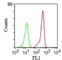
Western Blot 0.5-1ug/ml, Flow Cytometry 0.5-1ug/million cells, ELISA 1-5ug/ml for coating, Immunocytochemistry/Immunofluorescence 1-2ug/ml, Immunoprecipitation 1-2ug/500ug protein lysate, Immunohistochemistry-Paraffin 0.5-1ug/ml, Immunohistochemistry-Frozen 0.5-1ug/ml
Gene Accession No.
P16150
Gene Symbols
SPN
Immunogen
Myeloblastic KG1 cells were used as the immunogen for this antibody.
Quantity
0.1 mg
Research Discipline
B Cell Development and Differentiation Markers, Immunology
Gene ID (Entrez)
6693
Target Species
Human
Form
Purified
Applications
Western Blot, Flow Cytometry, ELISA, Immunocytochemistry, Immunofluorescence, Immunoprecipitation, Immunohistochemistry (Paraffin)
Clone
DF-T1
Conjugate
Unconjugated
Formulation
PBS with 0.05% BSA. with 0.05% Sodium Azide
Gene Alias
CD43 antigen, CD43), Galactoglycoprotein, GALGP, Leukocyte sialoglycoprotein, Sialophorin, sialophorin (gpL115, leukosialin, CD43)
Host Species
Mouse
Purification Method
Protein G purified
Regulatory Status
RUO
Primary or Secondary
Primary
Test Specificity
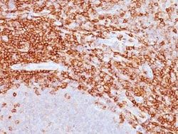
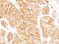
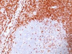
It recognizes a cell surface glycoprotein of 95/115/135kDa (depending upon the extent of glycosylation), identified as CD43. 70-90% of T-cell lymphomas and from 22-37% of B-cell lymphomas express CD43. No reactivity has been observed with reactive B-cells. So a B-lineage population that co-expresses CD43 is highly likely to be a malignant lymphoma, especially a low-grade lymphoma, rather than a reactive B-cell population. When CD43 antibody is used in combination with anti-CD20, effective immunophenotyping of the lymphomas in formalin-fixed tissues can be obtained. Co-staining of a lymphoid infiltrate with anti-CD20 and anti-CD43 argues against a reactive process and favors a diagnosis of lymphoma.
Content And Storage
Store at 4C.
Isotype
IgG1 κ
Related Products


Description
- CD43/Sialophorin Monoclonal specifically detects CD43/Sialophorin in Human samples
- It is validated for Western Blot, Flow Cytometry, Immunohistochemistry, Immunocytochemistry/Immunofluorescence, Immunohistochemistry-Paraffin.
Compare Similar Items
Show Difference
Antigen: CD43/Sialophorin
Classification: Monoclonal
Concentration: 0.2mg/mL
Dilution: Western Blot 0.5-1ug/ml, Flow Cytometry 0.5-1ug/million cells, ELISA 1-5ug/ml for coating, Immunocytochemistry/Immunofluorescence 1-2ug/ml, Immunoprecipitation 1-2ug/500ug protein lysate, Immunohistochemistry-Paraffin 0.5-1ug/ml, Immunohistochemistry-Frozen 0.5-1ug/ml
Gene Accession No.: P16150
Gene Symbols: SPN
Immunogen: Myeloblastic KG1 cells were used as the immunogen for this antibody.
Quantity: 0.1 mg
Research Discipline: B Cell Development and Differentiation Markers, Immunology
Gene ID (Entrez): 6693
Target Species: Human
Form: Purified
Applications: Western Blot, Flow Cytometry, ELISA, Immunocytochemistry, Immunofluorescence, Immunoprecipitation, Immunohistochemistry (Paraffin)
Clone: DF-T1
Conjugate: Unconjugated
Formulation: PBS with 0.05% BSA. with 0.05% Sodium Azide
Gene Alias: CD43 antigen, CD43), Galactoglycoprotein, GALGP, Leukocyte sialoglycoprotein, Sialophorin, sialophorin (gpL115, leukosialin, CD43)
Host Species: Mouse
Purification Method: Protein G purified
Regulatory Status: RUO
Primary or Secondary: Primary
Test Specificity: It recognizes a cell surface glycoprotein of 95/115/135kDa (depending upon the extent of glycosylation), identified as CD43. 70-90% of T-cell lymphomas and from 22-37% of B-cell lymphomas express CD43. No reactivity has been observed with reactive B-cells. So a B-lineage population that co-expresses CD43 is highly likely to be a malignant lymphoma, especially a low-grade lymphoma, rather than a reactive B-cell population. When CD43 antibody is used in combination with anti-CD20, effective immunophenotyping of the lymphomas in formalin-fixed tissues can be obtained. Co-staining of a lymphoid infiltrate with anti-CD20 and anti-CD43 argues against a reactive process and favors a diagnosis of lymphoma.
Content And Storage: Store at 4C.
Isotype: IgG1 κ
Antigen: CD43/Sialophorin
Classification: Monoclonal
Concentration: 0.2mg/mL
Dilution: Western Blot 0.5-1ug/ml, Flow Cytometry 0.5-1ug/million cells, ELISA 1-5ug/ml for coating, Immunocytochemistry/Immunofluorescence 1-2ug/ml, Immunoprecipitation 1-2ug/500ug protein lysate, Immunohistochemistry-Paraffin 0.5-1ug/ml, Immunohistochemistry-Frozen 0.5-1ug/ml
Gene Accession No.: P16150
Gene Symbols: SPN
Immunogen: Myeloblastic KG1 cells were used as the immunogen for this antibody.
Quantity: 0.2 mg
Research Discipline: B Cell Development and Differentiation Markers, Immunology
Gene ID (Entrez): 6693
Target Species: Human
Form: Purified
Applications: Western Blot, Flow Cytometry, ELISA, Immunocytochemistry, Immunofluorescence, Immunoprecipitation, Immunohistochemistry (Paraffin)
Clone: DF-T1
Conjugate: Unconjugated
Formulation: PBS with 0.05% BSA. with 0.05% Sodium Azide
Gene Alias: CD43 antigen, CD43), Galactoglycoprotein, GALGP, Leukocyte sialoglycoprotein, Sialophorin, sialophorin (gpL115, leukosialin, CD43)
Host Species: Mouse
Purification Method: Protein G purified
Regulatory Status: RUO
Primary or Secondary: Primary
Test Specificity: It recognizes a cell surface glycoprotein of 95/115/135kDa (depending upon the extent of glycosylation), identified as CD43. 70-90% of T-cell lymphomas and from 22-37% of B-cell lymphomas express CD43. No reactivity has been observed with reactive B-cells. So a B-lineage population that co-expresses CD43 is highly likely to be a malignant lymphoma, especially a low-grade lymphoma, rather than a reactive B-cell population. When CD43 antibody is used in combination with anti-CD20, effective immunophenotyping of the lymphomas in formalin-fixed tissues can be obtained. Co-staining of a lymphoid infiltrate with anti-CD20 and anti-CD43 argues against a reactive process and favors a diagnosis of lymphoma.
Content And Storage: Store at 4C.
Isotype: IgG1 κ
Antigen: Kappa Light Chain
Classification: Monoclonal
Concentration: 0.2mg/mL
Dilution: Western Blot 0.5-1ug/ml, Simple Western 10 ug/ml, Flow Cytometry 0.5-2ug/million cells, Immunohistochemistry-Paraffin 0.5-1ug/ml, Immunohistochemistry-Frozen 0.5-1ug/ml, SDS-Page
Gene Accession No.: P01601
Gene Symbols: IGKC
Immunogen: B lymphoma cells
Quantity: 0.1 mg
Research Discipline: __
Gene ID (Entrez): 3514
Target Species: Human
Form: Purified
Applications: Western Blot, Flow Cytometry, Immunohistochemistry (Paraffin), Immunohistochemistry (Frozen)
Clone: L1C1
Conjugate: Unconjugated
Formulation: PBS with 0.05% BSA. with 0.05% Sodium Azide
Gene Alias: HCAK1, IGKCD, immunoglobulin kappa constant, Km, MGC111575, MGC62011, MGC72072, MGC88770, MGC88771, MGC88809
Host Species: Mouse
Purification Method: Protein G purified
Regulatory Status: RUO
Primary or Secondary: Primary
Test Specificity: This MAb is specific to kappa light chain of immunoglobulin and shows no cross-reaction with lambda light chain or any of the five heavy chains. In mammals, the two light chains in an antibody are always identical, with only one type of light chain, kappa or lambda. The ratio of kappa to lambda is 70:30. However, with the occurrence of multiple myeloma or other B-cell malignancies this ratio is disturbed. Antibody to the kappa light chain is reportedly useful in the identification of leukemias, plasmacytomas, and certain non-Hodgkin's lymphomas. Demonstration of clonality in lymphoid infiltrates indicates that the infiltrate is malignant.
Content And Storage: Store at 4C.
Isotype: IgG1 κ