Kappa Light Chain Antibody (L1C1), Novus Biologicals™
Manufacturer: Fischer Scientific
Select a Size
| Pack Size | SKU | Availability | Price |
|---|---|---|---|
| Each of 1 | NBP21519102-Each-of-1 | In Stock | ₹ 56,159.00 |
NBP21519102 - Each of 1
In Stock
Quantity
1
Base Price: ₹ 56,159.00
GST (18%): ₹ 10,108.62
Total Price: ₹ 66,267.62
Antigen
Kappa Light Chain
Classification
Monoclonal
Concentration
0.2mg/mL
Dilution
Western Blot 0.5-1ug/ml, Simple Western 10 ug/ml, Flow Cytometry 0.5-2ug/million cells, Immunohistochemistry-Paraffin 0.5-1ug/ml, Immunohistochemistry-Frozen 0.5-1ug/ml, SDS-Page
Gene Accession No.
P01601
Gene Symbols
IGKC
Immunogen
B lymphoma cells
Purification Method
Protein G purified
Regulatory Status
RUO
Gene ID (Entrez)
3514
Target Species
Human
Form
Purified
Applications
Western Blot, Flow Cytometry, Immunohistochemistry (Paraffin), Immunohistochemistry (Frozen)
Clone
L1C1
Conjugate
Unconjugated
Formulation
PBS with 0.05% BSA. with 0.05% Sodium Azide
Gene Alias
HCAK1, IGKCD, immunoglobulin kappa constant, Km, MGC111575, MGC62011, MGC72072, MGC88770, MGC88771, MGC88809
Host Species
Mouse
Molecular Weight of Antigen
22.5 kDa
Quantity
0.2 mg
Primary or Secondary
Primary
Test Specificity
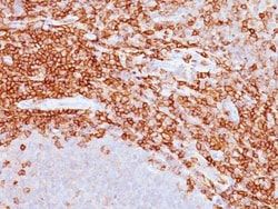
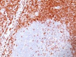
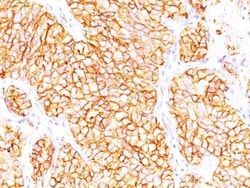
This MAb is specific to kappa light chain of immunoglobulin and shows no cross-reaction with lambda light chain or any of the five heavy chains. In mammals, the two light chains in an antibody are always identical, with only one type of light chain, kappa or lambda. The ratio of kappa to lambda is 70:30. However, with the occurrence of multiple myeloma or other B-cell malignancies this ratio is disturbed. Antibody to the kappa light chain is reportedly useful in the identification of leukemias, plasmacytomas, and certain non-Hodgkin's lymphomas. Demonstration of clonality in lymphoid infiltrates indicates that the infiltrate is malignant.
Content And Storage
Store at 4C.
Isotype
IgG1 κ
Related Products


Description
- Description Kappa Light Chain Monoclonal specifically detects Kappa Light Chain in Human samples
- It is validated for Western Blot, Simple Western, Immunohistochemistry, Immunohistochemistry-Paraffin.
Compare Similar Items
Show Difference

Antigen: Kappa Light Chain
Classification: Monoclonal
Concentration: 0.2mg/mL
Dilution: Western Blot 0.5-1ug/ml, Simple Western 10 ug/ml, Flow Cytometry 0.5-2ug/million cells, Immunohistochemistry-Paraffin 0.5-1ug/ml, Immunohistochemistry-Frozen 0.5-1ug/ml, SDS-Page
Gene Accession No.: P01601
Gene Symbols: IGKC
Immunogen: B lymphoma cells
Purification Method: Protein G purified
Regulatory Status: RUO
Gene ID (Entrez): 3514
Target Species: Human
Form: Purified
Applications: Western Blot, Flow Cytometry, Immunohistochemistry (Paraffin), Immunohistochemistry (Frozen)
Clone: L1C1
Conjugate: Unconjugated
Formulation: PBS with 0.05% BSA. with 0.05% Sodium Azide
Gene Alias: HCAK1, IGKCD, immunoglobulin kappa constant, Km, MGC111575, MGC62011, MGC72072, MGC88770, MGC88771, MGC88809
Host Species: Mouse
Molecular Weight of Antigen: 22.5 kDa
Quantity: 0.2 mg
Primary or Secondary: Primary
Test Specificity: This MAb is specific to kappa light chain of immunoglobulin and shows no cross-reaction with lambda light chain or any of the five heavy chains. In mammals, the two light chains in an antibody are always identical, with only one type of light chain, kappa or lambda. The ratio of kappa to lambda is 70:30. However, with the occurrence of multiple myeloma or other B-cell malignancies this ratio is disturbed. Antibody to the kappa light chain is reportedly useful in the identification of leukemias, plasmacytomas, and certain non-Hodgkin's lymphomas. Demonstration of clonality in lymphoid infiltrates indicates that the infiltrate is malignant.
Content And Storage: Store at 4C.
Isotype: IgG1 κ
Antigen: CD45RA
Classification: Monoclonal
Concentration: 0.2mg/mL
Dilution: Western Blot 0.5-1ug/ml, Flow Cytometry 0.5-1ug/million cells, ELISA 1-5ug/ml for coating, Immunocytochemistry/Immunofluorescence 1-2ug/ml, Immunoprecipitation 1-2ug/500ug protein lysate, Immunohistochemistry-Paraffin 0.5-1ug/ml, Immunohistochemistry-Frozen 0.5-1ug/ml
Gene Accession No.: P08575
Gene Symbols: PTPRC
Immunogen: Stimulated human leukocytes were used as immunogen to generate the CD45RA antibody.
Purification Method: Protein G purified
Regulatory Status: RUO
Gene ID (Entrez): 5788
Target Species: Human, Primate
Form: Purified
Applications: Western Blot, Flow Cytometry, ELISA, Immunocytochemistry, Immunofluorescence, Immunoprecipitation, Immunohistochemistry (Paraffin)
Clone: 158-4D3
Conjugate: Unconjugated
Formulation: PBS with 0.05% BSA. with 0.05% Sodium Azide
Gene Alias: B220, CD45 antigen, CD45R, EC 3.1.3.48, L-CA, LY5, protein tyrosine phosphatase, receptor type, C, receptor-type tyrosine-protein phosphatase C, T200 glycoprotein, T200 leukocyte common antigen, T200receptor type, c polypeptide
Host Species: Mouse
Molecular Weight of Antigen: __
Quantity: 0.1 mg
Primary or Secondary: Primary
Test Specificity: Recognizes a protein of 205kDa-220kDa, identified as CD45RA [Workshop V; Code CD45.38]. CD45RA is isoforms of the human leukocyte common antigen (CD45). Human CD45 contains three exons which encode peptide segments designated A, B and C, respectively. The differential splicing of the exons generates at least five isoforms, ABC, AB, BC, B and O. This antibody reacts with ABC and BC isoforms. CD45RA is expressed on 40-50% of peripheral CD4+ T-cells, 50% of peripheral CD8+ T-cells, B-cells, and leukemic B-cell lines. T-cells expressing CD45RA are naive or virgin T-cells. T-cells expressing CD45RO are memory T-cells. CD45RA and CD45RO define complementary, predominantly non-overlapping populations of resting peripheral T-cells. This MAb is useful in study on the subpopulation of CD4+ or CD8+ T-cells. It can especially be used to differentiate T-cell lymphomas (CD45RO +ve) from B cell lymphomas (CD45RA +ve).
Content And Storage: Store at 4C.
Isotype: IgG2a κ
Antigen: Mucin 5AC
Classification: Monoclonal
Concentration: 0.2mg/mL
Dilution: Flow Cytometry 0.5-1ug/million cells, Immunocytochemistry/Immunofluorescence 1-2ug/ml, Immunohistochemistry-Paraffin 0.5-1ug/ml, Immunohistochemistry-Frozen 0.5ug/ml, Flow (Intracellular)
Gene Accession No.: P98088
Gene Symbols: MUC5AC
Immunogen: M1 mucin preparation from the fluid of an ovarian mucinous cyst belonging to an O Le(a-b) patient
Purification Method: Protein G purified
Regulatory Status: RUO
Gene ID (Entrez): 4586
Target Species: Human, Mouse, Rat, Porcine, Chicken, Feline, Primate, Rabbit, Bovine (Negative)
Form: Purified
Applications: Flow Cytometry, Immunocytochemistry, Immunofluorescence, Immunohistochemistry (Paraffin), Immunohistochemistry (Frozen)
Clone: 45M1
Conjugate: Unconjugated
Formulation: PBS with 0.05% BSA. with 0.05% Sodium Azide
Gene Alias: gastric mucin, leB, lewis B blood group antigen, major airway glycoprotein, MUC5, mucin 5, subtypes A and C, tracheobronchial/gastric, mucin 5AC, oligomeric mucus/gel-forming, mucin 5AC, oligomeric mucus/gel-forming pseudogene, mucin-5 subtype AC, tracheobronchial, mucin-5AC, TBM, tracheobronchial mucin
Host Species: Mouse
Molecular Weight of Antigen: __
Quantity: 0.1 mg
Primary or Secondary: Primary
Test Specificity: This MAb recognizes the peptide core of gastric mucin M1 (>1,000kDa) (recently identified as Mucin 5AC). Its epitope is destroyed by beta-mercaptoethanol and proteases but not by periodate treatment. Antibody to gastric mucin M1 reacts with the gastric epithelium of normal human gastrointestinal tract as well as with the precancerous and cancerous colon but not with normal adult colon. It also reacts with fetal colonic mucosa. Resurgence of gastric mucin reactivity during colonic carcinogenesis is due to re-expression of the peptide core of gastric (or fetal colonic) mucins.
Content And Storage: Store at 4C.
Isotype: IgG1 κ