Laminin gamma 1 Antibody (SPM193), Novus Biologicals™
Rat Monoclonal Antibody
Manufacturer: Fischer Scientific
The price for this product is unavailable. Please request a quote
Antigen
Laminin gamma 1
Concentration
0.2mg/mL
Applications
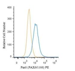
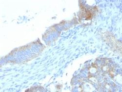
Flow Cytometry, Immunohistochemistry (Paraffin), Immunofluorescence
Conjugate
Unconjugated
Host Species
Rat
Research Discipline
Apoptosis, Cancer, Cytoskeleton Markers, Extracellular Matrix, Tumor Suppressors
Formulation
10mM PBS and 0.05% BSA with 0.05% Sodium Azide
Gene ID (Entrez)
3915
Immunogen
Murine EHS laminin preparation
Primary or Secondary
Primary
Content And Storage
Store at 4C.
Molecular Weight of Antigen
210 kDa
Clone
SPM193
Dilution
Flow Cytometry 0.5 - 1 ug/million cells in 0.1 ml, Immunohistochemistry-Paraffin 0.5 - 1.0 ug/ml, Immunofluorescence 1 - 2 ug/ml
Classification
Monoclonal
Form
Purified
Regulatory Status
RUO
Target Species
Human, Mouse
Gene Alias
LAMB2Laminin-7 subunit gamma, Laminin B2 chain, laminin subunit gamma-1, laminin, gamma 1 (formerly LAMB2), Laminin-1 subunit gamma, Laminin-10 subunit gamma, Laminin-11 subunit gamma, Laminin-2 subunit gamma, Laminin-3 subunit gamma, Laminin-4 subunit gamma, Laminin-6 subunit gamma, Laminin-8 subunit gamma, Laminin-9 subunit gamma, MGC87297, S-LAM gamma, S-laminin subunit gamma
Gene Symbols
LAMC1
Isotype
IgG2a κ
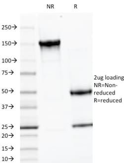
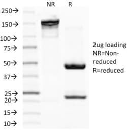
Purification Method
Protein A or G purified
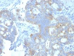
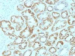
Test Specificity
Laminins are large hetero-trimeric, non-collagenous glycoproteins composed of alpha, beta, and gamma chains. This MAb reacts with laminin B2/1 chain of ∼210kDa and does not cross-react with other basement membrane components or fibronectin. Its specificity was established by immunoprecipitation and immunofluorescence of human skeletal muscle and kidney with laminin chain-specific MAbs. Epithelial sheets in vivo are separated from the mesenchymal elements of the stroma by a thin layer of a specialized type of extracellular matrix termed the basement membrane (BM). This structure consists of individual components, some of which are ubiquitous in BMs and some are not. The ubiquitous ones comprise laminin (LN), entactin/nidogen (EN), collagen type IV (CIV), and large heparan sulfate proteoglycan (HSPG), which interact specifically with each other to form a continuous and regular BM. Alterations of BM integrity, from local discontinuities up to complete loss, are described in many types
Related Products
Description
- Ensure accurate, reproducible results in Flow Cytometry, Immunohistochemistry (Paraffin), Immunofluorescence Laminin gamma 1 Monoclonal specifically detects Laminin gamma 1 in Human, Mouse samples
- It is validated for Flow Cytometry, Immunohistochemistry, Immunocytochemistry/Immunofluorescence, Immunohistochemistry-Paraffin, Immunofluorescence.
Compare Similar Items
Show Difference
Antigen: Laminin gamma 1
Concentration: 0.2mg/mL
Applications: Flow Cytometry, Immunohistochemistry (Paraffin), Immunofluorescence
Conjugate: Unconjugated
Host Species: Rat
Research Discipline: Apoptosis, Cancer, Cytoskeleton Markers, Extracellular Matrix, Tumor Suppressors
Formulation: 10mM PBS and 0.05% BSA with 0.05% Sodium Azide
Gene ID (Entrez): 3915
Immunogen: Murine EHS laminin preparation
Primary or Secondary: Primary
Content And Storage: Store at 4C.
Molecular Weight of Antigen: 210 kDa
Clone: SPM193
Dilution: Flow Cytometry 0.5 - 1 ug/million cells in 0.1 ml, Immunohistochemistry-Paraffin 0.5 - 1.0 ug/ml, Immunofluorescence 1 - 2 ug/ml
Classification: Monoclonal
Form: Purified
Regulatory Status: RUO
Target Species: Human, Mouse
Gene Alias: LAMB2Laminin-7 subunit gamma, Laminin B2 chain, laminin subunit gamma-1, laminin, gamma 1 (formerly LAMB2), Laminin-1 subunit gamma, Laminin-10 subunit gamma, Laminin-11 subunit gamma, Laminin-2 subunit gamma, Laminin-3 subunit gamma, Laminin-4 subunit gamma, Laminin-6 subunit gamma, Laminin-8 subunit gamma, Laminin-9 subunit gamma, MGC87297, S-LAM gamma, S-laminin subunit gamma
Gene Symbols: LAMC1
Isotype: IgG2a κ
Purification Method: Protein A or G purified
Test Specificity: Laminins are large hetero-trimeric, non-collagenous glycoproteins composed of alpha, beta, and gamma chains. This MAb reacts with laminin B2/1 chain of ∼210kDa and does not cross-react with other basement membrane components or fibronectin. Its specificity was established by immunoprecipitation and immunofluorescence of human skeletal muscle and kidney with laminin chain-specific MAbs. Epithelial sheets in vivo are separated from the mesenchymal elements of the stroma by a thin layer of a specialized type of extracellular matrix termed the basement membrane (BM). This structure consists of individual components, some of which are ubiquitous in BMs and some are not. The ubiquitous ones comprise laminin (LN), entactin/nidogen (EN), collagen type IV (CIV), and large heparan sulfate proteoglycan (HSPG), which interact specifically with each other to form a continuous and regular BM. Alterations of BM integrity, from local discontinuities up to complete loss, are described in many types
Antigen: Ornithine Decarboxylase
Concentration: 0.2mg/mL
Applications: Western Blot, Flow Cytometry, Immunohistochemistry (Paraffin), SDS-Page, Immunofluorescence
Conjugate: Unconjugated
Host Species: Mouse
Research Discipline: Cell Cycle and Replication
Formulation: 10mM PBS and 0.05% BSA with 0.05% Sodium Azide
Gene ID (Entrez): 4953
Immunogen: Recombinant human ODC-1 protein
Primary or Secondary: Primary
Content And Storage: Store at 4C.
Molecular Weight of Antigen: 53 kDa
Clone: ODC1/487
Dilution: Western Blot 0.5 - 1.0 ug/ml, Flow Cytometry 0.5 - 1 ug/million cells in 0.1 ml, Immunohistochemistry-Paraffin 0.5 - 1.0 ug/ml, SDS-Page, Immunofluorescence 0.5 - 1.0 ug/ml
Classification: Monoclonal
Form: Purified
Regulatory Status: RUO
Target Species: Human, Rat
Gene Alias: ODCEC 4.1.1.17, ornithine decarboxylase, ornithine decarboxylase 1
Gene Symbols: ODC1
Isotype: IgG2a κ
Purification Method: Protein A or G purified
Test Specificity: Recognizes a 53kDa protein, identified as the Ornithine Decarboxylase (ODC-1). ODC is the initial and rate-limiting enzyme in the biosynthetic pathway of polyamines and is involved in the conversion of ornithine to putrescine. The biological activity of ODC-1 is rapidly induced in response to virtually all agents known to promote cell proliferation including hormones, drugs, growth factors, mitogens, and tumor promoters. Reportedly, ODC mRNA levels are elevated in lung carcinomas as well as in colon adenomas and carcinomas. ODC activity in colorectal carcinomas is greater than those in adenomas and normal mucosa.
