CDw17 Antibody (HO18.3G-6.F5), Novus Biologicals™
Mouse Monoclonal Antibody
Manufacturer: Fischer Scientific
The price for this product is unavailable. Please request a quote
Antigen
CDw17
Concentration
0.2 mg/mL
Applications
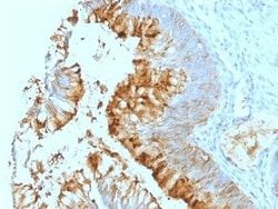
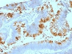
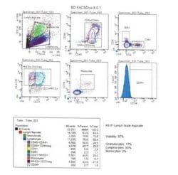
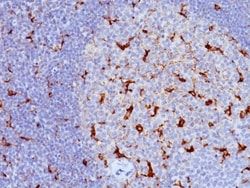
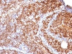
Flow Cytometry, Immunofluorescence
Conjugate
Unconjugated
Host Species
Mouse
Target Species
Human
Gene ID (Entrez)
8869
Immunogen
B-2 Microglobulin associated proteins from a detergent lysate of human PBL
Primary or Secondary
Primary
Content And Storage
Store at 4C.
Clone
HO18.3G-6.F5
Dilution
Flow Cytometry 0.5 - 1 ug/million cells in 0.1 ml, Immunofluorescence 0.5 - 1.0 ug/ml
Classification
Monoclonal
Form
Purified
Regulatory Status
RUO
Formulation
10mM PBS and 0.05% BSA with 0.05% Sodium Azide
Gene Symbols
ST3GAL5
Isotype
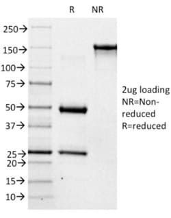
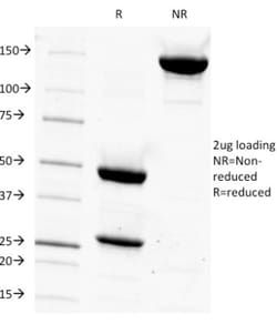
IgM
Purification Method
Protein A or G purified
Test Specificity
CD17 is an intermediate glycosphingolipid from the metabolism of higher gangliosides that localizes to sphingolipid-sterol rafts. CD17 is detectable in monocytes, granulocytes, basophils, platelets, a subset of peripheral B cells (CD19+) and tonsil dendritic cells. It is rapidly down regulated on activated granulocytes and is upregulated on IL-2 activated T lymphocytes. CD17 binds to bacteria and may function in phagocytosis. VEGF-treated endothelial cells can produce CD17, which can then mediate signaling toward PECAM-1 expression and angiogenesis. Tumor necrosis factor alpha (TNFalpha)-induced astrogliosis (astrocyte proliferation and glial fibrillary acidic protein (GFAP) upregulation) in response to neuro-inflammation (i.e. spinal cord injury) causes an increase in intracellular levels of CD17. Aberrant levels of glycosphingolipids are a feature of cancer cells and may influence integrin clustering and internalization.
Related Products

Description
- Ensure accurate, reproducible results in Flow Cytometry, Immunofluorescence CDw17 Monoclonal specifically detects CDw17 in Human samples
- It is validated for Flow Cytometry, Immunocytochemistry/Immunofluorescence, Immunofluorescence.
Compare Similar Items
Show Difference
Antigen: CDw17
Concentration: 0.2 mg/mL
Applications: Flow Cytometry, Immunofluorescence
Conjugate: Unconjugated
Host Species: Mouse
Target Species: Human
Gene ID (Entrez): 8869
Immunogen: B-2 Microglobulin associated proteins from a detergent lysate of human PBL
Primary or Secondary: Primary
Content And Storage: Store at 4C.
Clone: HO18.3G-6.F5
Dilution: Flow Cytometry 0.5 - 1 ug/million cells in 0.1 ml, Immunofluorescence 0.5 - 1.0 ug/ml
Classification: Monoclonal
Form: Purified
Regulatory Status: RUO
Formulation: 10mM PBS and 0.05% BSA with 0.05% Sodium Azide
Gene Symbols: ST3GAL5
Isotype: IgM
Purification Method: Protein A or G purified
Test Specificity: CD17 is an intermediate glycosphingolipid from the metabolism of higher gangliosides that localizes to sphingolipid-sterol rafts. CD17 is detectable in monocytes, granulocytes, basophils, platelets, a subset of peripheral B cells (CD19+) and tonsil dendritic cells. It is rapidly down regulated on activated granulocytes and is upregulated on IL-2 activated T lymphocytes. CD17 binds to bacteria and may function in phagocytosis. VEGF-treated endothelial cells can produce CD17, which can then mediate signaling toward PECAM-1 expression and angiogenesis. Tumor necrosis factor alpha (TNFalpha)-induced astrogliosis (astrocyte proliferation and glial fibrillary acidic protein (GFAP) upregulation) in response to neuro-inflammation (i.e. spinal cord injury) causes an increase in intracellular levels of CD17. Aberrant levels of glycosphingolipids are a feature of cancer cells and may influence integrin clustering and internalization.

Antigen: SUMO1
Concentration: 0.2mg/mL
Applications: Western Blot, Flow Cytometry, Immunohistochemistry (Paraffin), Immunofluorescence
Conjugate: Unconjugated
Host Species: Mouse
Target Species: Human, Rat
Gene ID (Entrez): 7341
Immunogen: Recombinant human SUMO1 protein
Primary or Secondary: Primary
Content And Storage: Store at 4C.
Clone: SUMO1/1188
Dilution: Western Blot 0.5 - 1.0 ug/ml, Flow Cytometry 0.5 - 1 ug/million cells in 0.1 ml, Immunohistochemistry-Paraffin 0.5 - 1.0 ug/ml, Immunofluorescence 0.5 - 1.0 ug/ml
Classification: Monoclonal
Form: Purified
Regulatory Status: RUO
Formulation: 10mM PBS and 0.05% BSA with 0.05% Sodium Azide
Gene Symbols: SUMO1
Isotype: IgG1 κ
Purification Method: Protein A or G purified
Test Specificity: This MAb is specific to SUMO-1 and shows no cross-reaction with either SUMO-2 or SUMO-3. The small ubiquitin-related modifier (SUMO) proteins, which include SUMO-1, SUMO-2 and SUMO-3, belong to the ubiquitin-like protein family. Like ubiquitin, the SUMO proteins are synthesized as precursor proteins that undergo processing before conjugation to target proteins. Also, both utilize the E1, E2, and E3 cascade enzymes for conjugation. However, SUMO and ubiquitin differ with respect to targeting. Ubiquitination predominantly targets proteins for degradation, whereas sumoylation targets proteins to a variety of cellular processing, including nuclear transport, transcriptional regulation, apoptosis and protein stability. The unconjugated SUMO-1 protein localizes to the nuclear membrane.
Antigen: SUMO2
Concentration: 0.2mg/mL
Applications: Flow Cytometry, Immunohistochemistry (Paraffin), Immunofluorescence
Conjugate: Unconjugated
Host Species: Mouse
Target Species: Human, Rat
Gene ID (Entrez): 6613
Immunogen: Recombinant human SUMO2 protein
Primary or Secondary: Primary
Content And Storage: Store at 4C.
Clone: SUMO2/1199
Dilution: Flow Cytometry 0.5 - 1 ug/million cells in 0.1 ml, Immunohistochemistry-Paraffin 0.5 - 1.0 ug/ml, Immunofluorescence 0.5 - 1.0 ug/ml
Classification: Monoclonal
Form: Purified
Regulatory Status: RUO
Formulation: 10mM PBS and 0.05% BSA with 0.05% Sodium Azide
Gene Symbols: SUMO2
Isotype: IgG1 κ
Purification Method: Protein A or G purified
Test Specificity: The small ubiquitin-related modifier (SUMO) proteins, which include SUMO-1, 2 and 3, belong to the ubiquitin-like protein family. Like ubiquitin, the SUMO proteins are synthesized as precursor proteins that undergo processing before conjugation to target proteins. Also, both utilize the E1, E2 and E3 cascade enzymes for conjugation. However, SUMO and ubiquitin differ with respect to targeting. Ubiquitination predominantly targets proteins for degradation, whereas sumoylation targets proteins to a variety of cellular processing, including nuclear transport, transcriptional regulation, apoptosis and protein stability. The unconjugated SUMO-1, 2 and 3 proteins localize to the nuclear membrane, nuclear bodies and cytoplasm, respectively. SUMO-1 utilizes Ubc9 for conjugation to several target proteins, which include MDM2, p53, PML and RanGap1. SUMO-2 and 3 contribute to a greater percentage of protein modification than does SUMO-1 and unlike SUMO-1, they can form polymeric chains. In additi