Lamin A/C, Mouse anti-Human, Monkey, Hamster, Mouse, Clone: 2A1, Millipore Sigma™
Manufacturer: Fischer Scientific
Select a Size
| Pack Size | SKU | Availability | Price |
|---|---|---|---|
| Each of 1 | MABT134025-Each-of-1 | In Stock | ₹ 17,118.26 |
MABT134025 - Each of 1
In Stock
Quantity
1
Base Price: ₹ 17,118.26
GST (18%): ₹ 3,081.287
Total Price: ₹ 20,199.547
Antigen
Lamin A/C
Classification
Monoclonal
Conjugate
Unconjugated
Formulation
Purified mouse monoclonal antibody IgG2b in buffer containing 0.1 M Tris-Glycine (pH 7.4), 150 mM NaCl with 0.05% sodium azide.
Gene Symbols
LMNA;LMN1
Immunogen
Purified protein corresponding to the rod domain for human Lamin A/C.
Quantity
25 μg
Research Discipline
Cell Structure
Gene ID (Entrez)
NP_733821
Target Species
Human, Monkey, Hamster, Mouse
Form
Purified
Applications
Immunocytochemistry, Immunofluorescence, Immunoprecipitation, Western Blot
Clone
2A1
Dilution
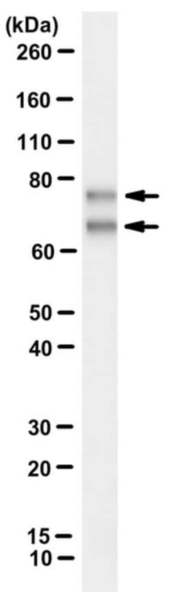
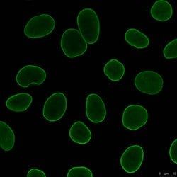
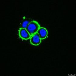
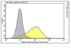
Immunoprecipitation Analysis: A representative immunoprecipitated Lamin A/C in culture supernatant (Data courtesy of Marie Lang, M.D., Stefan Schuchner, Ph.D. and Egon Ogris, M.D., Medical University of Vienna, Austria). Western Blotting Analysis: A representative lot detected Lamin A/C in culture supernatants of various cell lines (Data courtesy of Marie Lang, M.D., Stefan Schuchner, Ph.D. and Egon Ogris, M.D., Medical University of Vienna, Austria). Immunocytochemistry Analysis: A 1:1,000 dilution from a representative lot detected Lamin A/C in HeLa cells. Immunofluorescence Analysis: A representative lot detected Lamin A/C in the nuclear interior of HeLa cells (Data courtesy of Marie Lang, M.D., Stefan Schuchner, Ph.D. and Egon Ogris, M.D., Medical University of Vienna, Austria).
Gene Alias
70 kDa Lamin;Renal carcinoma antigen NY-REN-32
Host Species
Mouse
Purification Method
Protein G purified
Regulatory Status
RUO
Primary or Secondary
Primary
Test Specificity
Clone 2A1 detects the Lamin A/C species in the nuclear interior in multiple species. It targets the Rod domain of Lamin A/C.
Content And Storage
Stable for 1 year at 2-8°C from date of receipt.
Isotype
IgG2b κ
Related Products

Description
- Anti-Lamin A/C, clone 2A1, Cat
- No
- MABT1340, is a highly specific mouse monoclonal antibody that targets Lamin A/C and has been tested for use in Immunocytochemistry, Immunofluorescence, Immunoprecipitation, and Western Blotting
- Prelamin-A/C (UniProt: P02545) is encoded by the LMNA (also known as LMN1) gene (Gene ID: 4000) in human
- It is cleaved into Lamin-A/C (also known as 70 kDa Lamin, Renal carcinoma antigen NY-REN-32)
- Lamins are components of the nuclear lamina that provide a framework for the nuclear envelope and may also interact with chromatin
- Lamin A and C are present in equal amounts in the lamina of mammals
- Plays an important role in nuclear assembly, chromatin organization, nuclear membrane and telomere dynamics
- Lamin A is initially synthesized as prelamin A that undergoes several modifications in the carboxyl terminal region that allow incorporation of prelamin A into the nuclear envelope and its subsequent processing into the mature lamin A
- Cleavage of 15 residues (aa 647-662) by ZMPSTE24/FACE1 generates the final protein product
- Unlike mature lamin A, prelamin A accumulates as discrete and localized foci at the nuclear periphery
- Prelamin-A/C can accelerate smooth muscle cell senescence
- It can act to disrupt mitosis and induce DNA damage in vascular smooth muscle cells (VSMCs), leading to mitotic failure, genomic instability, and premature senescence
- Mutations in LMNA gene are known to cause Emery-Dreifuss muscular dystrophy that is characterized by weakness and atrophy of muscle without involvement of the nervous system
- Some mutations have also been linked to familial type of lipodystrophy characterized by the loss of subcutaneous adipose tissue in the lower parts of the body
- (Ref.: Casasola, A., et al
- (2016)
- Nucleus 7(1); 84-102).
Compare Similar Items
Show Difference
Antigen: Lamin A/C
Classification: Monoclonal
Conjugate: Unconjugated
Formulation: Purified mouse monoclonal antibody IgG2b in buffer containing 0.1 M Tris-Glycine (pH 7.4), 150 mM NaCl with 0.05% sodium azide.
Gene Symbols: LMNA;LMN1
Immunogen: Purified protein corresponding to the rod domain for human Lamin A/C.
Quantity: 25 μg
Research Discipline: Cell Structure
Gene ID (Entrez): NP_733821
Target Species: Human, Monkey, Hamster, Mouse
Form: Purified
Applications: Immunocytochemistry, Immunofluorescence, Immunoprecipitation, Western Blot
Clone: 2A1
Dilution: Immunoprecipitation Analysis: A representative immunoprecipitated Lamin A/C in culture supernatant (Data courtesy of Marie Lang, M.D., Stefan Schuchner, Ph.D. and Egon Ogris, M.D., Medical University of Vienna, Austria). Western Blotting Analysis: A representative lot detected Lamin A/C in culture supernatants of various cell lines (Data courtesy of Marie Lang, M.D., Stefan Schuchner, Ph.D. and Egon Ogris, M.D., Medical University of Vienna, Austria). Immunocytochemistry Analysis: A 1:1,000 dilution from a representative lot detected Lamin A/C in HeLa cells. Immunofluorescence Analysis: A representative lot detected Lamin A/C in the nuclear interior of HeLa cells (Data courtesy of Marie Lang, M.D., Stefan Schuchner, Ph.D. and Egon Ogris, M.D., Medical University of Vienna, Austria).
Gene Alias: 70 kDa Lamin;Renal carcinoma antigen NY-REN-32
Host Species: Mouse
Purification Method: Protein G purified
Regulatory Status: RUO
Primary or Secondary: Primary
Test Specificity: Clone 2A1 detects the Lamin A/C species in the nuclear interior in multiple species. It targets the Rod domain of Lamin A/C.
Content And Storage: Stable for 1 year at 2-8°C from date of receipt.
Isotype: IgG2b κ
Antigen: Lamin A/C
Classification: Monoclonal
Conjugate: Unconjugated
Formulation: Purified mouse monoclonal antibody IgG2a in buffer containing 0.1 M Tris-Glycine (pH 7.4), 150 mM NaCl with 0.05% sodium azide.
Gene Symbols: LMNA;LMN1
Immunogen: Purified protein corresponding to the Ig-fold of human Lamin A/C.
Quantity: 100 μg
Research Discipline: Cell Structure
Gene ID (Entrez): NP_001244303
Target Species: Monkey, Syrian Hamster, Rat, Mouse, Human
Form: Purified
Applications: ChIP Assay, Immunofluorescence, Immunoprecipitation, Western Blot
Clone: 4C11
Dilution: Western Blotting Analysis: A representative lot detected Lamin A/C in Western Blotting applications (Gesson, K., et. al. (2016). Genome Res. 26(4):462-73; Roblek, M., et. al. (2010). PLoS One. 5(5):e10604).Dot Blot Analysis: A representative lot detected Lamin A/C in Dot Blot applications (Roblek, M., et. al. (2010). PLoS One. 5(5):e10604). Immunocytochemistry Analysis: A 1:1,000 dilution from a representative lot detected Lamin A/C in HeLa cells.Western Blotting Analysis: A representative lot detected Lamin A/C in WB used to demonstrate high titration and excellent properties of clone 4C11 (Courtesy of Marie lang, M.D., Stefan Schuchner, Ph.D. and Egon Ogris, M.D., Medical University of Vienna, Austria).Immunofluorescence Analysis: A representative lot detected the Lamin A/C species at the nuclear lamina of HeLa cells (Courtesy of Marie Lang, M.D., Stefan Schuchner, Ph.D. and Egon Ogris, M.D., Medical University of Vienna, Austria).Immunoprecipitation Analysis: A representative lot de
Gene Alias: 70 kDa Lamin;Renal carcinoma antigen NY-REN-32
Host Species: Mouse
Purification Method: Protein G purified
Regulatory Status: RUO
Primary or Secondary: Primary
Test Specificity: Clone 4C11 detects Lamin A/C at the nuclear lamina in multiple species.
Content And Storage: Stable for 1 year at 2-8°C from date of receipt.
Isotype: IgG2a κ
Antigen: Lamin A/C
Classification: Monoclonal
Conjugate: Unconjugated
Formulation: Purified mouse monoclonal antibody IgG2a in buffer containing 0.1 M Tris-Glycine (pH 7.4), 150 mM NaCl with 0.05% sodium azide.
Gene Symbols: LMNA;LMN1
Immunogen: Purified protein corresponding to the Ig-fold of human Lamin A/C.
Quantity: 25 μg
Research Discipline: Cell Structure
Gene ID (Entrez): NP_001244303
Target Species: Monkey, Syrian Hamster, Rat, Mouse, Human
Form: Purified
Applications: ChIP Assay, Immunofluorescence, Immunoprecipitation, Western Blot
Clone: 4C11
Dilution: Western Blotting Analysis: A representative lot detected Lamin A/C in Western Blotting applications (Gesson, K., et. al. (2016). Genome Res. 26(4):462-73; Roblek, M., et. al. (2010). PLoS One. 5(5):e10604).Dot Blot Analysis: A representative lot detected Lamin A/C in Dot Blot applications (Roblek, M., et. al. (2010). PLoS One. 5(5):e10604). Immunocytochemistry Analysis: A 1:1,000 dilution from a representative lot detected Lamin A/C in HeLa cells.Western Blotting Analysis: A representative lot detected Lamin A/C in WB used to demonstrate high titration and excellent properties of clone 4C11 (Courtesy of Marie lang, M.D., Stefan Schuchner, Ph.D. and Egon Ogris, M.D., Medical University of Vienna, Austria).Immunofluorescence Analysis: A representative lot detected the Lamin A/C species at the nuclear lamina of HeLa cells (Courtesy of Marie Lang, M.D., Stefan Schuchner, Ph.D. and Egon Ogris, M.D., Medical University of Vienna, Austria).Immunoprecipitation Analysis: A representative lot de
Gene Alias: 70 kDa Lamin;Renal carcinoma antigen NY-REN-32
Host Species: Mouse
Purification Method: Protein G purified
Regulatory Status: RUO
Primary or Secondary: Primary
Test Specificity: Clone 4C11 detects Lamin A/C at the nuclear lamina in multiple species.
Content And Storage: Stable for 1 year at 2-8°C from date of receipt.
Isotype: IgG2a κ