Aminopeptidase N (CD13), Mouse anti-Human, Clone: 452, Millipore Sigma™
Mouse Monoclonal Antibody
Manufacturer: Fischer Scientific
The price for this product is unavailable. Please request a quote
Antigen
Aminopeptidase N (CD13)
Dilution
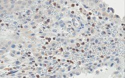
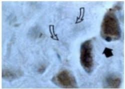
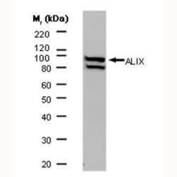
Immunocytochemistry Analysis: A representative lot detected Aminopeptidase N (CD13) in Immunocytochemistry applications (Rahman, M.M., et. al. (2014). Front Physiol. 4:402; Subramani, J., et. al. (2013). J Immunol. 191(7):3905-12).Flow Cytometry Analysis: A representative lot detected Aminopeptidase N (CD13) in Flow Cytometry applications (Subramani, J., et. al. (2013). J Immunol. 191(7):3905-12).Western Blotting Analysis: A representative lot detected Aminopeptidase N (CD13) in Western Blotting applications (Rahman, M.M., et. al. (2014). Stem Cells. 32(6):1564-77; Rahman, M.M., et. al. (2014). Front Physiol. 4:402).Agonist Analysis: A representative lot induced a temporal increase in phosphorylation of FAK, ERK and Src kinases in U937 cells. (Subramani, J., et. al. (2013). J Immunol. 191(7):3905-12).Immunoprecipitation Analysis: A representative lot immunoprecipitated Aminopeptidase N (CD13) in Immunoprecipitation applications (Subramani, J., et. al. (2013). J Immunol. 191(7):3905-12)
Classification
Monoclonal
Form
Purified
Regulatory Status
RUO
Target Species
Human
Gene Alias
AP-N;hAPN;Alanyl aminopeptidase;Aminopeptidase M;AP-M;Microsomal aminopeptidase;Myeloid plasma membrane glycoprotein CD13;gp150;CD13
Gene Symbols
ANPEP;APN;CD13;PEPN
Isotype
IgG1 κ
Purification Method
Protein G purified
Test Specificity
Clone 452 specifically detects Aminopeptidase N (CD13) in human cells.
Clone
452
Applications
Flow Cytometry, Immunocytochemistry, Immunoprecipitation, Inhibition Assays, Western Blot
Conjugate
Unconjugated
Host Species
Mouse
Research Discipline
Inflammation & Immunology
Formulation
Purified mouse monoclonal antibody IgG1 in PBS without azide.
Gene ID (Entrez)
NP_001141
Immunogen
Isolated human dermal fibroblasts.
Primary or Secondary
Primary
Content And Storage
Stable for 1 year at -20°C from date of receipt. Handling Recommendations: Upon receipt and prior to removing the cap, centrifuge the vial and gently mix the solution. Aliquot into microcentrifuge tubes and store at -20°C. Avoid repeated freeze/thaw cycles, which may damage IgG and affect product performance.
Related Products


Description
- Anti-Aminopeptidase N (CD13), clone 452, Cat
- No
- MABF2147, is a mouse monoclonal antibody that detects Aminopeptidase N and has been tested for use in Flow Cytometry, Immunocytochemistry, Immunoprecipitation, Agonist function, and Western Blotting
- Aminopeptidase N (UniProt: P15144; also known as EC:3.4.11.2, AP-N, hAPN, Alanyl aminopeptidase, Aminopeptidase M, AP-M, Microsomal aminopeptidase, Myeloid plasma membrane glycoprotein CD13, gp150, CD13) is encoded by the ANPEP (also known as APN, CD13, PEPN) gene (Gene ID: 290) in human
- Aminopeptidase N is a single-pass type II membrane protein that is also found as a soluble form in cells
- It is widely expressed as a homodimer of 280 kDa on the cell surface in many tissues, including intestinal epithelia and the nervous system
- It serves as a broad specificity aminopeptidase that plays a role in the final digestion of peptides generated from hydrolysis of proteins by gastric and pancreatic proteases
- It preferentially cleaves N-terminus neutral amino acids, most notably alanine residue
- Aminopeptidase N is involved in many physiological processes, including antigen presentation regulation, differentiation, proliferation, apoptosis, cancer metastasis, and angiogenesis
- It is also involved in the processing of various peptides including peptide hormones, such as angiotensin III and IV, neuropeptides, and chemokines
- Human Aminopeptidase N consists of a short N-terminal cytoplasmic end (aa 2-8), a transmembrane domain (aa 9-32), and a large extracellular portion (aa 33-967), which is composed of a Ser/Thr-rich region (aa 33-68) and the metalloprotease domain (aa 69-967).
Compare Similar Items
Show Difference

Antigen: Aminopeptidase N (CD13)
Dilution: Immunocytochemistry Analysis: A representative lot detected Aminopeptidase N (CD13) in Immunocytochemistry applications (Rahman, M.M., et. al. (2014). Front Physiol. 4:402; Subramani, J., et. al. (2013). J Immunol. 191(7):3905-12).Flow Cytometry Analysis: A representative lot detected Aminopeptidase N (CD13) in Flow Cytometry applications (Subramani, J., et. al. (2013). J Immunol. 191(7):3905-12).Western Blotting Analysis: A representative lot detected Aminopeptidase N (CD13) in Western Blotting applications (Rahman, M.M., et. al. (2014). Stem Cells. 32(6):1564-77; Rahman, M.M., et. al. (2014). Front Physiol. 4:402).Agonist Analysis: A representative lot induced a temporal increase in phosphorylation of FAK, ERK and Src kinases in U937 cells. (Subramani, J., et. al. (2013). J Immunol. 191(7):3905-12).Immunoprecipitation Analysis: A representative lot immunoprecipitated Aminopeptidase N (CD13) in Immunoprecipitation applications (Subramani, J., et. al. (2013). J Immunol. 191(7):3905-12)
Classification: Monoclonal
Form: Purified
Regulatory Status: RUO
Target Species: Human
Gene Alias: AP-N;hAPN;Alanyl aminopeptidase;Aminopeptidase M;AP-M;Microsomal aminopeptidase;Myeloid plasma membrane glycoprotein CD13;gp150;CD13
Gene Symbols: ANPEP;APN;CD13;PEPN
Isotype: IgG1 κ
Purification Method: Protein G purified
Test Specificity: Clone 452 specifically detects Aminopeptidase N (CD13) in human cells.
Clone: 452
Applications: Flow Cytometry, Immunocytochemistry, Immunoprecipitation, Inhibition Assays, Western Blot
Conjugate: Unconjugated
Host Species: Mouse
Research Discipline: Inflammation & Immunology
Formulation: Purified mouse monoclonal antibody IgG1 in PBS without azide.
Gene ID (Entrez): NP_001141
Immunogen: Isolated human dermal fibroblasts.
Primary or Secondary: Primary
Content And Storage: Stable for 1 year at -20°C from date of receipt. Handling Recommendations: Upon receipt and prior to removing the cap, centrifuge the vial and gently mix the solution. Aliquot into microcentrifuge tubes and store at -20°C. Avoid repeated freeze/thaw cycles, which may damage IgG and affect product performance.

Antigen: TMPRSS2
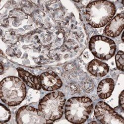
Dilution: Immunohistochemistry (Paraffin) Analysis: 1:50 dilution from a representative lot detected TMPRSS2 in human kidney tissue sections.Enzyme Immunoassay (ELISA) Analysis: A representative lot detected TMPRSS2 in ELISA applications (Lucas, J.M., et. al. (2008). J Pathol. 215(2):118-25).Western Blotting Analysis: A representative lot detected TMPRSS2 in Western Blotting applications (Lucas, J.M., et. al. (2008). J Pathol. 215(2):118-25).Immunohistochemistry (Paraffin) Analysis: A representative lot detected TMPRSS2 in Immunohistochemistry applications (Lucas, J.M., et. al. (2008). J Pathol. 215(2):118-25; Bertram, S., et. al. (2012). PLoS One. 7(4):e35876).
Classification: Monoclonal
Form: Purified
Regulatory Status: RUO
Target Species: Human
Gene Alias: Transmembrane protease serine 2;Serine protease 10
Gene Symbols: TMPRSS2;PRSS10
Isotype: IgG1 κ
Purification Method: Protein G purified
Test Specificity: Clone P5H9-A3 specifically detects human Transmembrane protease serine 2. It targets an epitope with in the extracellular serine protease domain.
Clone: P5H9-A3
Applications: ELISA, Immunohistochemistry (Paraffin), Western Blot
Conjugate: Unconjugated
Host Species: Mouse
Research Discipline: Inflammation & Immunology
Formulation: Purified mouse monoclonal antibody IgG1 in buffer containing 0.1 M Tris-Glycine (pH 7.4), 150 mM NaCl with 0.05% sodium azide.
Gene ID (Entrez): __
Immunogen: KLH-conjugated linear peptide corresponding to 16 amino acids from the extracellular domain of human transmembrane protease serine 2.
Primary or Secondary: Primary
Content And Storage: Stable for 1 year at 2-8°C from date of receipt.

Antigen: CD63 (LAMP3)
Dilution: Immunohistochemistry (Paraffin) Analysis: A 1:250 dilution from a representative lot detected CD63 (LAMP3) in human spleen and human bone marrow tissue sections.Immunocytochemistry Analysis: A representative lot detected CD63 (LAMP3) in Immunocytochemistry applications (Atkinson, B., et. al. (1984). Cancer Res. 44(6):2577-81).Flow Cytometry Analysis: A representative lot detected CD63 (LAMP3) in Flow Cytometry applications (Li, J., et. al. (2003). J Immunol. 171(6):2922-9).Western Blotting Analysis: A representative lot detected CD63 (LAMP3) in Western Blotting applications (Smith, M., et. al. (1997). Melanoma Res. 7 Suppl 2:S163-70).Immunohistochemistry Analysis: A representative lot detected CD63 (LAMP3) in Immunohistochemistry applications (Li, J., et. al. (2003). J Immunol. 171(6):2922-9).
Classification: Monoclonal
Form: Purified
Regulatory Status: RUO
Target Species: Human
Gene Alias: CD63 antigen;Granulophysin;Lysosomal-associated membrane protein 3;LAMP-3;Melanoma-associated antigen ME491;OMA81H;Ocular melanoma-associated antigen;Tetraspanin-30;Tspan-30
Gene Symbols: CD63;MLA1;TSPAN30
Isotype: IgG1 κ
Purification Method: Protein G purified
Test Specificity: Clone ME491 specifically detects CD63 (LAMP-3) in human cells.
Clone: ME491
Applications: Flow Cytometry, Immunocytochemistry, Immunohistochemistry (Paraffin), Western Blot
Conjugate: Unconjugated
Host Species: Mouse
Research Discipline: Inflammation & Immunology
Formulation: Purified mouse monoclonal antibody IgG1 in buffer containing 0.1 M Tris-Glycine (pH 7.4), 150 mM NaCl with 0.05% sodium azide.
Gene ID (Entrez): NP_001244318
Immunogen: Clear supernatant from SK-Mel-23 cell lysate.
Primary or Secondary: Primary
Content And Storage: Stable for 1 year at 2-8°C from date of receipt.