MilliporeSigma™ APP, CT, Ascites Free, Mouse, Unlabeled, Clone: 2.F2.19B4,
Manufacturer: MilliporeSigma™
Select a Size
| Pack Size | SKU | Availability | Price |
|---|---|---|---|
| Each of 1 | MAB343CMI-Each-of-1 | In Stock | ₹ 40,633.84 |
MAB343CMI - Each of 1
In Stock
Quantity
1
Base Price: ₹ 40,633.84
GST (18%): ₹ 7,314.091
Total Price: ₹ 47,947.931
Antigen
APP, CT, Ascites Free
Classification
Monoclonal
Concentration
Please refer to lot specific datasheet.
Formulation
Purified mouse monoclonal IgG1κ antibody in buffer containing 0.1M Tris-Glycine (pH 7.4), 150mM NaCl with 0.05% Sodium Azide.
Gene Accession No.
P05067
Immunogen
APP C-terminal 53-a.a. fragment.
Quantity
100 μg
Research Discipline
Neuroscience
Gene ID (Entrez)
NP_000475
Target Species
Human
Form
Purified
Applications
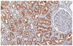
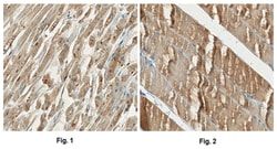
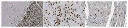
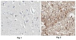
Immunocytochemistry, Immunofluorescence, Immunohistochemistry (Paraffin), Immunoprecipitation, Western Blot
Clone
2.F2.19B4
Conjugate
Unconjugated
Gene
APP, A4, AD1
Host Species
Mouse
Purification Method
Protein G Purified
Regulatory Status
RUO
Primary or Secondary
Primary
Test Specificity
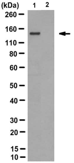
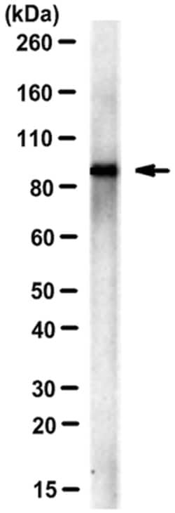
Clone 2.F2.19B4, also known as the Jonas antibody, regcognizes a C-terminal epitope present in 10 of the 11 human spliced isoforms reported by UniProt (P05067), with APP305 being the only spliced isoform lacking the targeted epitope. Clone 2.F2.19B4 recognizes full-length APP, the APP C-terminal fragments (CTFs) produced by α- and β-secretase cleavage, and the APP intracellular domain (AICD) generated by γ-secretase cleavage, but not the sAPPα or sAPPβ fragment. Epitope has been mapped to APP770 a.a. 732-751, which is equivalent to APP695 a.a. 657-676 (Van Vickle, G.D., et al. (2007). Biochemistry. 46(36):10317-10327).
Content And Storage
Stable for 1 year at 2°-8°C from date of receipt.
Isotype
IgG1 κ
Related Products


Description
- Amyloid beta A4 protein (UniProt P05067; also known as ABPP, Alzheimer disease amyloid protein, Amyloid precursor protein, APP, APPI, Cerebral vascular amyloid peptide, CVAP, PN-II, PreA4, Protease nexin-II) is encoded by the APP (also known as A4, AD1) gene (Gene ID 351) in human
- Amyloid precursor protein (APP) is initially produced with a signal peptide sequence (a.a
- 1-17), the removal of which yields the mature protein with a large extracellular portion (a.a
- 18-699), followed by a transmembrane segment (a.a
- 700-723) and a cytoplasmic (a.a
- 724-770) tail
- APP can be further processed by the α-, β-, and γ-secretases in two alternative processing pathways
- In the non-amyloidogenic pathway, APP is first cleaved by the plasma membrane-localized α-secretase to generate an N-terminal extracellular sAPPα fragment (a.a
- 18-687) and a membrane-bound C-terminal fragment C83 (CTFα), which can be further cleaved by γ-secretase to produce a non-toxic small peptide p3 and a cytoplasmic APP intracellular domain (AICD)
- In the amyloidogenic pathway, APP undergoes β-cleavage in BACE-1 (β-site APP-cleaving enzyme)-enriched endosomes to generate an N-terminal extracellular sAPPβ fragment (a.a
- 18-671) and a membrane-bound C-terminal fragment C99 (CTFβ)
- Subsequent cleavage of C99 by γ-secretase releases the amyloid β peptides, Aβ1-42 (672-713) and Aβ1-40 (672-711), and AICD
- Aβ accumulation in the cortical and hippocampal regions of the brain is a major pathological feature of Alzheimer's disease (AD)
- Aβ Ser8 phosphorylation is shown to promote Aβ aggregation into oligomeric and fibrillar assemblies and to prevent Aβ proteolytic clearance by certain proteases.
Compare Similar Items
Show Difference
Antigen: APP, CT, Ascites Free
Classification: Monoclonal
Concentration: Please refer to lot specific datasheet.
Formulation: Purified mouse monoclonal IgG1κ antibody in buffer containing 0.1M Tris-Glycine (pH 7.4), 150mM NaCl with 0.05% Sodium Azide.
Gene Accession No.: P05067
Immunogen: APP C-terminal 53-a.a. fragment.
Quantity: 100 μg
Research Discipline: Neuroscience
Gene ID (Entrez): NP_000475
Target Species: Human
Form: Purified
Applications: Immunocytochemistry, Immunofluorescence, Immunohistochemistry (Paraffin), Immunoprecipitation, Western Blot
Clone: 2.F2.19B4
Conjugate: Unconjugated
Gene: APP, A4, AD1
Host Species: Mouse
Purification Method: Protein G Purified
Regulatory Status: RUO
Primary or Secondary: Primary
Test Specificity: Clone 2.F2.19B4, also known as the Jonas antibody, regcognizes a C-terminal epitope present in 10 of the 11 human spliced isoforms reported by UniProt (P05067), with APP305 being the only spliced isoform lacking the targeted epitope. Clone 2.F2.19B4 recognizes full-length APP, the APP C-terminal fragments (CTFs) produced by α- and β-secretase cleavage, and the APP intracellular domain (AICD) generated by γ-secretase cleavage, but not the sAPPα or sAPPβ fragment. Epitope has been mapped to APP770 a.a. 732-751, which is equivalent to APP695 a.a. 657-676 (Van Vickle, G.D., et al. (2007). Biochemistry. 46(36):10317-10327).
Content And Storage: Stable for 1 year at 2°-8°C from date of receipt.
Isotype: IgG1 κ

Antigen: APP
Classification: Monoclonal
Concentration: __
Formulation: Liquid.
Gene Accession No.: NM_000484.2; NM_201413.1; NM_201414.1
Immunogen: Carboxyl fragment of APP 643-695 / Jonas.
Quantity: 100 μL
Research Discipline: __
Gene ID (Entrez): __
Target Species: Human, Rat
Form: Ascites
Applications: Immunohistochemistry (Paraffin), Western Blot
Clone: 2.F2.19B4
Conjugate: Unconjugated
Gene: __
Host Species: Mouse
Purification Method: Unpurified
Regulatory Status: __
Primary or Secondary: Primary
Test Specificity: __
Content And Storage: −20°C in undiluted aliquots for up to 6 months. Avoid repeated freeze/thaw cycles.
Isotype: IgG1

Antigen: Endothelin-1
Classification: Monoclonal
Concentration: LYOPH
Formulation: Lyophilized from a 0.2 μm filtered solution in PBS with Trehalose. *Small pack size (SP) is supplied as a 0.2 μm filtered solution in PBS. with No Preservative
Gene Accession No.: __
Immunogen: Human Endothelin-1
Quantity: 100 μg
Research Discipline: __
Gene ID (Entrez): 1906
Target Species: Human
Form: Purified
Applications: ELISA, Immunocytochemistry
Clone: 3G10
Conjugate: Unconjugated
Gene: __
Host Species: Rat
Purification Method: Protein A or G purified from hybridoma culture supernatant
Regulatory Status: RUO
Primary or Secondary: Primary
Test Specificity: Detects human Endothelin-1 C-Terminus. Recognizes the C-terminal region of mature Endothelin-1, -2, and -3 (amino acid residues 15-21).
Content And Storage: Use a manual defrost freezer and avoid repeated freeze-thaw cycles.12 months from date of receipt, -20 to -70 degreesC as supplied. 1 month, 2 to 8 degreesC under sterile conditions after reconstitution. 6 months, -20 to -70 degreesC under sterile conditions after reconstitution.
Isotype: IgG2b
