CD30/TNFRSF8 Antibody (CD30/412), Novus Biologicals™
Manufacturer: Novus Biologicals
Select a Size
| Pack Size | SKU | Availability | Price |
|---|---|---|---|
| Each of 1 | NBP229425-Each-of-1 | In Stock | ₹ 46,636.00 |
NBP229425 - Each of 1
In Stock
Quantity
1
Base Price: ₹ 46,636.00
GST (18%): ₹ 8,394.48
Total Price: ₹ 55,030.48
Antigen
CD30/TNFRSF8
Classification
Monoclonal
Concentration
0.2 mg/ml
Dilution
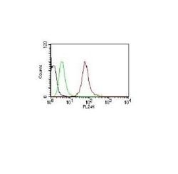
Flow Cytometry 0.5-1ug/million cells, ELISA 1-5ug/ml for coating, Immunocytochemistry/Immunofluorescence 1-2ug/ml, Immunohistochemistry-Paraffin 0.5-1.0ug/ml, Immunohistochemistry-Frozen 0.5-1.0ug/ml
Gene Alias
CD30, CD30 antigen, CD30KI-1, CD30L receptor, cytokine receptor CD30, D1S166EKi-1, Ki-1 antigen, Lymphocyte activation antigen CD30, tumor necrosis factor receptor superfamily member 8, tumor necrosis factor receptor superfamily, member 8
Host Species
Mouse
Purification Method
Protein A or G purified
Regulatory Status
RUO
Primary or Secondary
Primary
Test Specificity
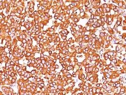
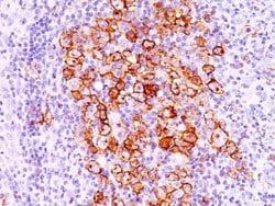
Recognizes a single chain glycoprotein of 105/120kDa, identified as CD30/Ki-1. CD30 is synthesized as a 90kDa precursor, which is processed in the Golgi complex into a membrane-bound phosphorylated mature 105/120kDa glycoprotein. In Hodgkins disease, CD30/Ki-1 antigen is expressed by mononuclear-Hodgkin and multinucleated Reed-Sternberg cells. It is also expressed by the tumor cells of a majority of anaplastic large cell lymphomas as well as by a varying proportion of activated T and B cells. This monoclonal antibody distinguishes large cell lymphomas derived from activated lymphoid cells from histiocytic malignancies and lymphomas derived from resting and precursor lymphoid cells or from anaplastic carcinomas. About one third of the Ki-1 positive lymphomas lack the leukocyte common antigen (CD45).
Content And Storage
Store at 4C.
Isotype
IgG1 κ
Applications
Flow Cytometry, ELISA, Immunocytochemistry, Immunofluorescence, Immunohistochemistry (Paraffin), Immunohistochemistry (Frozen)
Clone
CD30/412
Conjugate
Unconjugated
Gene Accession No.
P28908
Gene Symbols
TNFRSF8
Immunogen
Human CD30/TNFRSF8 recombinant protein (Uniprot: P28908)
Quantity
0.1 mg
Research Discipline
Apoptosis, Cell Cycle and Replication, Embryonic Stem Cell Markers, Immunology, Stem Cell Markers
Gene ID (Entrez)
943
Target Species
Human
Form
Purified
Related Products


Description
- CD30/TNFRSF8 Monoclonal specifically detects CD30/TNFRSF8 in Human samples
- It is validated for Immunohistochemistry, Immunohistochemistry-Paraffin.
Compare Similar Items
Show Difference
Antigen: CD30/TNFRSF8
Classification: Monoclonal
Concentration: 0.2 mg/ml
Dilution: Flow Cytometry 0.5-1ug/million cells, ELISA 1-5ug/ml for coating, Immunocytochemistry/Immunofluorescence 1-2ug/ml, Immunohistochemistry-Paraffin 0.5-1.0ug/ml, Immunohistochemistry-Frozen 0.5-1.0ug/ml
Gene Alias: CD30, CD30 antigen, CD30KI-1, CD30L receptor, cytokine receptor CD30, D1S166EKi-1, Ki-1 antigen, Lymphocyte activation antigen CD30, tumor necrosis factor receptor superfamily member 8, tumor necrosis factor receptor superfamily, member 8
Host Species: Mouse
Purification Method: Protein A or G purified
Regulatory Status: RUO
Primary or Secondary: Primary
Test Specificity: Recognizes a single chain glycoprotein of 105/120kDa, identified as CD30/Ki-1. CD30 is synthesized as a 90kDa precursor, which is processed in the Golgi complex into a membrane-bound phosphorylated mature 105/120kDa glycoprotein. In Hodgkins disease, CD30/Ki-1 antigen is expressed by mononuclear-Hodgkin and multinucleated Reed-Sternberg cells. It is also expressed by the tumor cells of a majority of anaplastic large cell lymphomas as well as by a varying proportion of activated T and B cells. This monoclonal antibody distinguishes large cell lymphomas derived from activated lymphoid cells from histiocytic malignancies and lymphomas derived from resting and precursor lymphoid cells or from anaplastic carcinomas. About one third of the Ki-1 positive lymphomas lack the leukocyte common antigen (CD45).
Content And Storage: Store at 4C.
Isotype: IgG1 κ
Applications: Flow Cytometry, ELISA, Immunocytochemistry, Immunofluorescence, Immunohistochemistry (Paraffin), Immunohistochemistry (Frozen)
Clone: CD30/412
Conjugate: Unconjugated
Gene Accession No.: P28908
Gene Symbols: TNFRSF8
Immunogen: Human CD30/TNFRSF8 recombinant protein (Uniprot: P28908)
Quantity: 0.1 mg
Research Discipline: Apoptosis, Cell Cycle and Replication, Embryonic Stem Cell Markers, Immunology, Stem Cell Markers
Gene ID (Entrez): 943
Target Species: Human
Form: Purified
Antigen: pan Cytokeratin
Classification: Monoclonal
Concentration: 0.2 mg/ml
Dilution: Western Blot 1 - 2 ug/mL, Flow Cytometry 1 - 2 ug/ million cells, Immunohistochemistry, Immunocytochemistry/Immunofluorescence 1 - 2 ug/mL, Immunohistochemistry-Paraffin 0.25 - 1.0 ug/mL, Immunohistochemistry-Frozen 0.25 - 1.0 ug/mL, Flow (Intracellular), CyTOF-reported, Single Cell Western 1:10, Dual RNAscope ISH-IHC
Gene Alias: __
Host Species: Mouse
Purification Method: Protein A or G purified
Regulatory Status: RUO
Primary or Secondary: Primary
Test Specificity: Twenty human keratins are resolved with two-dimensional gel electrophoresis into acidic (pI 6.0) subfamilies. This antibody cocktail recognizes acidic (Type I or LMW) and basic (Type II or HMW) cytokeratins, which 67kDa (CK1); 64kDa (CK3); 59kDa (CK4); 58kDa (CK5); 56kDa (CK6); 52kDa (CK8); 56.5kDa (CK10); 50kDa (CK14); 50kDa (CK15); 48kDa (CK16); 40kDa (CK19). Many studies have shown the usefulness of keratins as markers in cancer research and tumor diagnosis. AE-1/AE-3 is a broad spectrum anti pan-cytokeratin antibody cocktail, which differentiates epithelial tumors from non-epithelial tumors e.g. squamous vs. adenocarcinoma of the lung, liver carcinoma, breast cancer, and esophageal cancer. It has been used to characterize the source of various neoplasms and to study the distribution of cytokeratin containing cells in epithelia during normal development and during the development of epithelial neoplasms. This antibody stains cytokeratins present in normal and abnormal human tissues
Content And Storage: Store at 4C.
Isotype: IgG1 κ
Applications: Western Blot, Flow Cytometry, Immunohistochemistry, Immunocytochemistry, Immunofluorescence, Immunohistochemistry (Paraffin), Immunohistochemistry (Frozen), CyTOF, Western Blot, In Situ Hybridization (ISH)
Clone: AE-1/AE-3
Conjugate: Unconjugated
Gene Accession No.: Q7Z794, Q01546
Gene Symbols: KRT1
Immunogen: This Cytokeratin, pan Antibody (AE-1/AE-3) was developed against total keratin isolated from human epidermal callus was used as immunogen to generate the pan cytokeratin antibodies AE1 + AE3 (Woodcock-Mitchell, 1982).
Quantity: 0.1 mg
Research Discipline: Apoptosis, Cancer, Cell Biology, Cellular Markers, Signal Transduction
Gene ID (Entrez): 3848
Target Species: Human, Mouse, Rat, Bovine, Canine, Chicken, Primate, Rabbit, Reptile, Zebrafish
Form: Purified
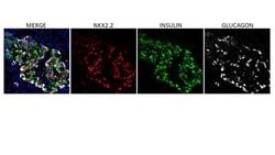
Antigen: NKX2.2
Classification: Monoclonal
Concentration: 0.2 mg/mL
Dilution: Flow Cytometry 0.5-1ug/million cells, Immunocytochemistry/Immunofluorescence 1-2ug/ml, Immunohistochemistry-Paraffin 0.5-1.0ug/ml, Immunohistochemistry-Frozen 0.5-1.0ug/ml
Gene Alias: Homeobox protein NK-2 homolog B, NK-2 (Drosophila) homolog B, NK2 homeobox 2, NK2 transcription factor related, locus 2, NK2 transcription factor related, locus 2 (Drosophila), NK2 transcription factor-like protein B, Nkx2.2, NKX2.2homeobox protein Nkx-2.2, NKX2BNK-2 homolog B
Host Species: Mouse
Purification Method: Protein A or G purified
Regulatory Status: RUO
Primary or Secondary: Primary
Test Specificity: Expression of NKX2.2 has been found in neuroendocrine tumors of the gut, making it a potential marker for the study of gastrointestinal neuroendocrine tumors. More recently, NKX2.2 protein was identified as a target of EWS-FLI-1, the fusion protein specific to Ewing sarcoma, and was shown to be differentially upregulated in Ewing sarcoma on the basis of array-based gene expression analysis. It acts as a valuable marker for Ewing sarcoma, with a sensitivity of 93% and a specificity of 89%, and aids in the differential diagnosis of small round cell tumors.
Content And Storage: Store at 4C.
Isotype: IgG2b κ
Applications: Flow Cytometry, Immunocytochemistry, Immunofluorescence, Immunohistochemistry (Paraffin), Immunohistochemistry (Frozen)
Clone: NX2/294
Conjugate: Unconjugated
Gene Accession No.: O95096
Gene Symbols: NKX2-2
Immunogen: Human full-length recombinant NKX2.2 protein (Uniprot: O95096)
Quantity: 0.1 mg
Research Discipline: __
Gene ID (Entrez): 4821
Target Species: Human, Mouse, Rat, Chicken
Form: Purified