p53 Antibody (BP53-12), Novus Biologicals™
Manufacturer: Novus Biologicals
Select a Size
| Pack Size | SKU | Availability | Price |
|---|---|---|---|
| Each of 1 | NBP229453-Each-of-1 | In Stock | ₹ 47,704.00 |
NBP229453 - Each of 1
In Stock
Quantity
1
Base Price: ₹ 47,704.00
GST (18%): ₹ 8,586.72
Total Price: ₹ 56,290.72
Antigen
p53
Classification
Monoclonal
Concentration
0.2 mg/ml
Dilution
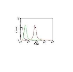
Western Blot 0.5-1ug/ml, Simple Western 10 ug/ml, Flow Cytometry 0.5-1ug/million cells in 0.1ml, Immunocytochemistry/Immunofluorescence 1-2ug/ml, Immunoprecipitation 1-2ug/500ug protein lysate, Immunohistochemistry-Paraffin 0.5-1.0ug/ml, Immunohistochemistry-Frozen 0.5-1.0ug/ml
Gene Alias
Antigen NY-CO-13, FLJ92943, LFS1TRP53, p53, p53 tumor suppressor, P53cellular tumor antigen p53, Phosphoprotein p53, transformation-related protein 53, tumor protein p53, Tumor suppressor p53
Host Species
Mouse
Molecular Weight of Antigen
53 kDa
Quantity
0.1 mg
Research Discipline
Apoptosis, Cancer, Cell Cycle and Replication, Cellular Markers, Checkpoint signaling, Core ESC Like Genes, DNA Double Strand Break Repair, DNA Repair, HIF Target Genes, Hypoxia, Neuroscience, Neurotransmission, p53 Pathway, Phospho Specific, Stem Cell Markers, Transcription Factors and Regulators, Tumor Suppressors
Gene ID (Entrez)
7157
Target Species
Human, Mouse, Canine, Chicken, Hamster, Monkey, Mouse (Negative), Rat (Negative)
Form
Purified
Applications
Western Blot, Flow Cytometry, Immunocytochemistry, Immunofluorescence, Immunoprecipitation
Clone
BP53-12
Conjugate
Unconjugated
Gene Accession No.
P04637
Gene Symbols
TP53
Immunogen
Recombinant human wild-type p53 protein (Uniprot: P04637)
Purification Method
Protein A or G purified
Regulatory Status
RUO
Primary or Secondary
Primary
Test Specificity
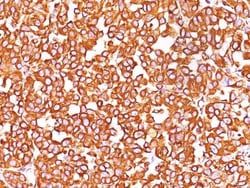
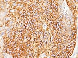
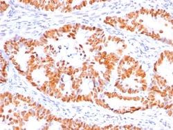
This monoclonal antibody reacts with an N-terminal epitope (aa 16-25) of both wild type and mutated p53. Mutation and/or allelic loss of p53 is one of the causes of a variety of mesenchymal and epithelial tumors. If it occurs in the germ line, such tumors run in families. In most transformed and tumor cells the concentration of p53 is increased 51000 fold over the minute concentrations (1000 molecules cell) in normal cells, principally due to the increased half-life (4 h) compared to that of the wild-type (20 min). p53 Localizes in the nucleus, but is detectable at the plasma membrane during mitosis and when certain mutations modulate cytoplasmic/nuclear distribution. Mutations arise with an average frequency of 70% but incidence varies from zero in carcinoid lung tumors to 97% in primary melanomas. High concentrations of p53 protein are transiently expressed in human epidermis and superficial dermal fibroblasts following mild ultraviolet irradiation. Positive nuclear staining with p53
Content And Storage
Store at 4C.
Isotype
IgG2a κ
Related Products


Description
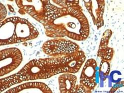
- Description p53 Monoclonal specifically detects p53 in Human, Canine, Chicken, Hamster, Monkey, Mouse (Negative), Rat (Negative) samples
- It is validated for Western Blot, Simple Western, Immunohistochemistry, Immunohistochemistry-Paraffin.
Compare Similar Items
Show Difference
Antigen: p53
Classification: Monoclonal
Concentration: 0.2 mg/ml
Dilution: Western Blot 0.5-1ug/ml, Simple Western 10 ug/ml, Flow Cytometry 0.5-1ug/million cells in 0.1ml, Immunocytochemistry/Immunofluorescence 1-2ug/ml, Immunoprecipitation 1-2ug/500ug protein lysate, Immunohistochemistry-Paraffin 0.5-1.0ug/ml, Immunohistochemistry-Frozen 0.5-1.0ug/ml
Gene Alias: Antigen NY-CO-13, FLJ92943, LFS1TRP53, p53, p53 tumor suppressor, P53cellular tumor antigen p53, Phosphoprotein p53, transformation-related protein 53, tumor protein p53, Tumor suppressor p53
Host Species: Mouse
Molecular Weight of Antigen: 53 kDa
Quantity: 0.1 mg
Research Discipline: Apoptosis, Cancer, Cell Cycle and Replication, Cellular Markers, Checkpoint signaling, Core ESC Like Genes, DNA Double Strand Break Repair, DNA Repair, HIF Target Genes, Hypoxia, Neuroscience, Neurotransmission, p53 Pathway, Phospho Specific, Stem Cell Markers, Transcription Factors and Regulators, Tumor Suppressors
Gene ID (Entrez): 7157
Target Species: Human, Mouse, Canine, Chicken, Hamster, Monkey, Mouse (Negative), Rat (Negative)
Form: Purified
Applications: Western Blot, Flow Cytometry, Immunocytochemistry, Immunofluorescence, Immunoprecipitation
Clone: BP53-12
Conjugate: Unconjugated
Gene Accession No.: P04637
Gene Symbols: TP53
Immunogen: Recombinant human wild-type p53 protein (Uniprot: P04637)
Purification Method: Protein A or G purified
Regulatory Status: RUO
Primary or Secondary: Primary
Test Specificity: This monoclonal antibody reacts with an N-terminal epitope (aa 16-25) of both wild type and mutated p53. Mutation and/or allelic loss of p53 is one of the causes of a variety of mesenchymal and epithelial tumors. If it occurs in the germ line, such tumors run in families. In most transformed and tumor cells the concentration of p53 is increased 51000 fold over the minute concentrations (1000 molecules cell) in normal cells, principally due to the increased half-life (4 h) compared to that of the wild-type (20 min). p53 Localizes in the nucleus, but is detectable at the plasma membrane during mitosis and when certain mutations modulate cytoplasmic/nuclear distribution. Mutations arise with an average frequency of 70% but incidence varies from zero in carcinoid lung tumors to 97% in primary melanomas. High concentrations of p53 protein are transiently expressed in human epidermis and superficial dermal fibroblasts following mild ultraviolet irradiation. Positive nuclear staining with p53
Content And Storage: Store at 4C.
Isotype: IgG2a κ
Antigen: Lambda Light Chain
Classification: Monoclonal
Concentration: 0.2 mg/ml
Dilution: Western Blot 0.5-1 μg/mL, Flow Cytometry 0.5-1 μg/million cells, ELISA 1-5 μg/mL for coating, Immunohistochemistry 0.5-1 μg/mL, Immunocytochemistry/Immunofluorescence 1-2 μg/mL, Immunoprecipitation 1-2 μg/500 μg protein lysate, Immunohistochemistry-Paraffin 0.5-1 μg/mL, Immunohistochemistry-Frozen 0.5-1 μg/mL
Gene Alias: IGLC, immunoglobulin lambda constant group
Host Species: Mouse
Molecular Weight of Antigen: 22.5 kDa
Quantity: 0.1 mg
Research Discipline: Adaptive Immunity, Immunology
Gene ID (Entrez): 3536
Target Species: Human
Form: Purified
Applications: Western Blot, Flow Cytometry, ELISA, Immunohistochemistry, Immunocytochemistry, Immunofluorescence, Immunoprecipitation, Immunohistochemistry (Paraffin), Immunohistochemistry (Frozen)
Clone: ICO-106
Conjugate: Unconjugated
Gene Accession No.: P01701, P01842
Gene Symbols: IGLC1
Immunogen: Human IgG was used as immunogen to generate the Lambda antibody.
Purification Method: Protein A or G purified
Regulatory Status: RUO
Primary or Secondary: Primary
Test Specificity: This monoclonal antibody is specific to lambda light chain of immunoglobulin and shows no cross-reaction with lambda light chain or any of the five heavy chains. In mammals, the two light chains in an antibody are always identical, with only one type of light chain, kappa or lambda. The ratio of Kappa to Lambda is 70:30. However, with the occurrence of multiple myeloma or other B-cell malignancies this ratio is disturbed. Antibody to the lambda light chain is reportedly useful in the identification of leukemias, plasmacytomas, and certain non-Hodgkins lymphomas. Demonstration of clonality in lymphoid infiltrates indicates that the infiltrate is malignant.
Content And Storage: Store at 4C.
Isotype: IgG1 κ

Antigen: p21/CIP1/CDKN1A
Classification: Monoclonal
Concentration: 0.2 mg/ml
Dilution: Western Blot, Flow Cytometry 0.5-1ug/million cells, Immunohistochemistry 0.5-1ug/ml, Immunocytochemistry/Immunofluorescence 1-2ug/ml, Immunohistochemistry-Paraffin 0.5-1ug/ml, Immunohistochemistry-Frozen 0.5-1ug/ml
Gene Alias: CAP20cyclin-dependent kinase inhibitor 1, CDK-interacting protein 1, CDKN1melanoma differentiation associated protein 6, CIP1WAF1CDK-interaction protein 1, cyclin-dependent kinase inhibitor 1A (p21, Cip1), MDA6, MDA-6, Melanoma differentiation-associated protein 6, p21, p21CIP1, p21Cip1/Waf1, PIC1, SDI1DNA synthesis inhibitor, wild-type p53-activated fragment 1
Host Species: Mouse
Molecular Weight of Antigen: 21 kDa
Quantity: 0.1 mg
Research Discipline: Apoptosis, Breast Cancer, Cancer, Cell Cycle and Replication, DNA Repair, Phospho Specific
Gene ID (Entrez): 1026
Target Species: Human, Mouse, Chimpanzee, Monkey
Form: Purified
Applications: Western Blot, Flow Cytometry, Immunohistochemistry, Immunocytochemistry, Immunofluorescence, Immunohistochemistry (Paraffin)
Clone: WA-1
Conjugate: Unconjugated
Gene Accession No.: P38936
Gene Symbols: CDKN1A
Immunogen: Human recombinant p21/CIP1/CDKN1A protein (Uniprot: P38936 )
Purification Method: Protein A or G purified
Regulatory Status: RUO
Primary or Secondary: Primary
Test Specificity: This monoclonal antibody recognizes a 21kDa protein, identified as the p21WAF1 tumor suppressor protein. This monoclonal antibody is highly specific to p21 and shows no cross-reaction with other closely related mitotic inhibitors. p21WAF1 is a specific inhibitor of cdks and a tumor suppressor involved in the pathogenesis of a variety of malignancies. The expression of this gene acts as an inhibitor of the cell cycle during G1 phase and is tightly controlled by the tumor suppressor protein p53. Its expression is induced by the wild type, but not mutant, p53 suppressor protein. Normal cells generally display a rather intense nuclear p21 expression. Loss of p21 expression has been reported in many carcinomas (gastric carcinoma, non-small cell lung carcinoma, thyroid carcinoma). In ELISA, monoclonal antibody WA-1 is useful either as a solid phase or for detection of p21 protein.
Content And Storage: Store at 4C.
Isotype: IgG1 κ
