p21/CIP1/CDKN1A Antibody (WA-1), Novus Biologicals™
Manufacturer: Novus Biologicals
Select a Size
| Pack Size | SKU | Availability | Price |
|---|---|---|---|
| Each of 1 | NBP229463-Each-of-1 | In Stock | ₹ 47,882.00 |
NBP229463 - Each of 1
In Stock
Quantity
1
Base Price: ₹ 47,882.00
GST (18%): ₹ 8,618.76
Total Price: ₹ 56,500.76
Antigen
p21/CIP1/CDKN1A
Classification
Monoclonal
Concentration
0.2 mg/ml
Dilution
Western Blot, Flow Cytometry 0.5-1ug/million cells, Immunohistochemistry 0.5-1ug/ml, Immunocytochemistry/Immunofluorescence 1-2ug/ml, Immunohistochemistry-Paraffin 0.5-1ug/ml, Immunohistochemistry-Frozen 0.5-1ug/ml
Gene Alias
CAP20cyclin-dependent kinase inhibitor 1, CDK-interacting protein 1, CDKN1melanoma differentiation associated protein 6, CIP1WAF1CDK-interaction protein 1, cyclin-dependent kinase inhibitor 1A (p21, Cip1), MDA6, MDA-6, Melanoma differentiation-associated protein 6, p21, p21CIP1, p21Cip1/Waf1, PIC1, SDI1DNA synthesis inhibitor, wild-type p53-activated fragment 1
Host Species
Mouse
Molecular Weight of Antigen
21 kDa
Quantity
0.1 mg
Research Discipline
Apoptosis, Breast Cancer, Cancer, Cell Cycle and Replication, DNA Repair, Phospho Specific
Gene ID (Entrez)
1026
Target Species
Human, Mouse, Chimpanzee, Monkey
Form
Purified
Applications
Western Blot, Flow Cytometry, Immunohistochemistry, Immunocytochemistry, Immunofluorescence, Immunohistochemistry (Paraffin)
Clone
WA-1
Conjugate
Unconjugated
Gene Accession No.
P38936
Gene Symbols
CDKN1A
Immunogen
Human recombinant p21/CIP1/CDKN1A protein (Uniprot: P38936 )
Purification Method
Protein A or G purified
Regulatory Status
RUO
Primary or Secondary
Primary
Test Specificity
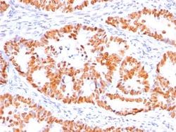
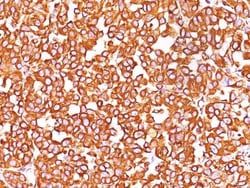
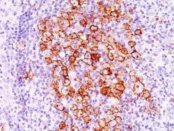
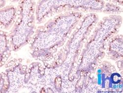
This monoclonal antibody recognizes a 21kDa protein, identified as the p21WAF1 tumor suppressor protein. This monoclonal antibody is highly specific to p21 and shows no cross-reaction with other closely related mitotic inhibitors. p21WAF1 is a specific inhibitor of cdks and a tumor suppressor involved in the pathogenesis of a variety of malignancies. The expression of this gene acts as an inhibitor of the cell cycle during G1 phase and is tightly controlled by the tumor suppressor protein p53. Its expression is induced by the wild type, but not mutant, p53 suppressor protein. Normal cells generally display a rather intense nuclear p21 expression. Loss of p21 expression has been reported in many carcinomas (gastric carcinoma, non-small cell lung carcinoma, thyroid carcinoma). In ELISA, monoclonal antibody WA-1 is useful either as a solid phase or for detection of p21 protein.
Content And Storage
Store at 4C.
Isotype
IgG1 κ
Related Products


Description
- Description p21/CIP1/CDKN1A Monoclonal antibody specifically detects p21/CIP1/CDKN1A in Human, Mouse, Chimpanzee, Monkey samples
- It is validated for Western Blot, Flow Cytometry, Immunohistochemistry, Immunocytochemistry, Immunofluorescence, Immunohistochemistry (Paraffin).
Compare Similar Items
Show Difference

Antigen: p21/CIP1/CDKN1A
Classification: Monoclonal
Concentration: 0.2 mg/ml
Dilution: Western Blot, Flow Cytometry 0.5-1ug/million cells, Immunohistochemistry 0.5-1ug/ml, Immunocytochemistry/Immunofluorescence 1-2ug/ml, Immunohistochemistry-Paraffin 0.5-1ug/ml, Immunohistochemistry-Frozen 0.5-1ug/ml
Gene Alias: CAP20cyclin-dependent kinase inhibitor 1, CDK-interacting protein 1, CDKN1melanoma differentiation associated protein 6, CIP1WAF1CDK-interaction protein 1, cyclin-dependent kinase inhibitor 1A (p21, Cip1), MDA6, MDA-6, Melanoma differentiation-associated protein 6, p21, p21CIP1, p21Cip1/Waf1, PIC1, SDI1DNA synthesis inhibitor, wild-type p53-activated fragment 1
Host Species: Mouse
Molecular Weight of Antigen: 21 kDa
Quantity: 0.1 mg
Research Discipline: Apoptosis, Breast Cancer, Cancer, Cell Cycle and Replication, DNA Repair, Phospho Specific
Gene ID (Entrez): 1026
Target Species: Human, Mouse, Chimpanzee, Monkey
Form: Purified
Applications: Western Blot, Flow Cytometry, Immunohistochemistry, Immunocytochemistry, Immunofluorescence, Immunohistochemistry (Paraffin)
Clone: WA-1
Conjugate: Unconjugated
Gene Accession No.: P38936
Gene Symbols: CDKN1A
Immunogen: Human recombinant p21/CIP1/CDKN1A protein (Uniprot: P38936 )
Purification Method: Protein A or G purified
Regulatory Status: RUO
Primary or Secondary: Primary
Test Specificity: This monoclonal antibody recognizes a 21kDa protein, identified as the p21WAF1 tumor suppressor protein. This monoclonal antibody is highly specific to p21 and shows no cross-reaction with other closely related mitotic inhibitors. p21WAF1 is a specific inhibitor of cdks and a tumor suppressor involved in the pathogenesis of a variety of malignancies. The expression of this gene acts as an inhibitor of the cell cycle during G1 phase and is tightly controlled by the tumor suppressor protein p53. Its expression is induced by the wild type, but not mutant, p53 suppressor protein. Normal cells generally display a rather intense nuclear p21 expression. Loss of p21 expression has been reported in many carcinomas (gastric carcinoma, non-small cell lung carcinoma, thyroid carcinoma). In ELISA, monoclonal antibody WA-1 is useful either as a solid phase or for detection of p21 protein.
Content And Storage: Store at 4C.
Isotype: IgG1 κ

Antigen: TIMP-3
Classification: Polyclonal
Concentration: 0.2 mg/ml
Dilution: Flow Cytometry 0.5-1ug/million cells, Immunohistochemistry 1-2ug/ml, Immunocytochemistry/Immunofluorescence 1-2ug/mlimmunohistochemistry 1-2ug/ml, Immunohistochemistry-Paraffin
Gene Alias: HSMRK222, K222, K222TA2, metalloproteinase inhibitor 3, MIG-5 protein, Protein MIG-5, pseudoinflammatory), SFD, TIMP metallopeptidase inhibitor 3, TIMP-3, tissue inhibitor of metalloproteinase 3 (Sorsby fundus dystrophy, Tissue inhibitor of metalloproteinases 3
Host Species: Rabbit
Molecular Weight of Antigen: 30 kDa
Quantity: 0.1 mg
Research Discipline: Angiogenesis, Apoptosis, Cancer, Hypoxia, Metastasis, Vision
Gene ID (Entrez): 7078
Target Species: Human, Bovine, Canine, Equine, Sheep
Form: Purified
Applications: Flow Cytometry, Immunohistochemistry, Immunocytochemistry, Immunofluorescence, Immunohistochemistry (Paraffin)
Clone: __
Conjugate: Unconjugated
Gene Accession No.: P35625
Gene Symbols: TIMP3
Immunogen: TIMP-3 (aa175-211) (Uniprot: P35625)
Purification Method: Protein A purified
Regulatory Status: RUO
Primary or Secondary: Primary
Test Specificity: TIMP3 (tissue inhibitor of metalloproteinases 3), along with family members TIMP1, TIMP2, and TIMP4, are inhibitors of the matrix metalloproteinases (MMPs), a group of peptidases involved in degradation of the extracellular matrix (ECM). An imbalance between MMPs and the associated TIMPs may play a significant role in the invasive phenotype of malignant tumors. TIMP s inhibit the proteolytic invasiveness of tumor cells and normal placental trophoblast cells. TIMP-3 may be involved in regulating trophoblastic invasion of the uterus as well as in regulating remodeling of the extracellular matrix during the folding of epithelia, and in the formation, branching and expansion of epithelial tubes.
Content And Storage: Store at 4C.
Isotype: IgG

Antigen: TIMP-3
Classification: Polyclonal
Concentration: 0.2mg/mL
Dilution: Flow Cytometry 0.5-1ug/million cells, Immunohistochemistry 1-2ug/ml, Immunocytochemistry/Immunofluorescence 1-2ug/mlimmunohistochemistry 1-2ug/ml, Immunohistochemistry-Paraffin
Gene Alias: HSMRK222, K222, K222TA2, metalloproteinase inhibitor 3, MIG-5 protein, Protein MIG-5, pseudoinflammatory), SFD, TIMP metallopeptidase inhibitor 3, TIMP-3, tissue inhibitor of metalloproteinase 3 (Sorsby fundus dystrophy, Tissue inhibitor of metalloproteinases 3
Host Species: Rabbit
Molecular Weight of Antigen: 30 kDa
Quantity: 0.2 mg
Research Discipline: Angiogenesis, Apoptosis, Cancer, Hypoxia, Metastasis, Vision
Gene ID (Entrez): 7078
Target Species: Human, Bovine, Canine, Equine, Sheep
Form: Purified
Applications: Flow Cytometry, Immunohistochemistry, Immunocytochemistry, Immunofluorescence, Immunohistochemistry (Paraffin)
Clone: __
Conjugate: Unconjugated
Gene Accession No.: P35625
Gene Symbols: TIMP3
Immunogen: A portion of amino acids 175-211 of human TIMP3 was used as immunogen for this antibody.
Purification Method: Protein A purified
Regulatory Status: RUO
Primary or Secondary: Primary
Test Specificity: TIMP3 (tissue inhibitor of metalloproteinases 3), along with family members TIMP1, TIMP2, and TIMP4, are inhibitors of the matrix metalloproteinases (MMPs), a group of peptidases involved in degradation of the extracellular matrix (ECM). An imbalance between MMPs and the associated TIMPs may play a significant role in the invasive phenotype of malignant tumors. TIMP s inhibit the proteolytic invasiveness of tumor cells and normal placental trophoblast cells. TIMP-3 may be involved in regulating trophoblastic invasion of the uterus as well as in regulating remodeling of the extracellular matrix during the folding of epithelia, and in the formation, branching and expansion of epithelial tubes.
Content And Storage: Store at 4C.
Isotype: IgG