UCH-L1/PGP9.5 Antibody (UCHL1/841), Novus Biologicals™
Mouse Monoclonal Antibody
Manufacturer: Fischer Scientific
The price for this product is unavailable. Please request a quote
Antigen
UCH-L1/PGP9.5
Concentration
0.2 mg/mL
Applications
Western Blot, Immunofluorescence
Conjugate
Unconjugated
Host Species
Mouse
Research Discipline
Alzheimers Research, Cellular Markers, Neurodegeneration, Neuronal Cell Markers, Neuroscience, Neurotransmission
Formulation
10mM PBS and 0.05% BSA with 0.05% Sodium Azide
Gene ID (Entrez)
7345
Immunogen
Recombinant human UCHL1 protein
Primary or Secondary
Primary
Content And Storage
Store at 4C.
Clone
UCHL1/841
Dilution
Western Blot 0.5 - 1.0 ug/ml, Immunofluorescence 1 - 2 ug/ml
Classification
Monoclonal
Form
Purified
Regulatory Status
RUO
Target Species
Human
Gene Alias
EC 3.4.19.12, EC 6.-, Neuron cytoplasmic protein 9.5, PARK5, PGP 9.5, PGP9.5, PGP9.5Uch-L1, PGP95, ubiquitin carboxyl-terminal esterase L1 (ubiquitin thiolesterase), ubiquitin carboxyl-terminal hydrolase isozyme L1, ubiquitin C-terminal hydrolase, Ubiquitin thioesterase L1, UCHL1, UCH-L1
Gene Symbols
UCHL1
Isotype
IgG2a κ
Purification Method
Protein A or G purified
Test Specificity
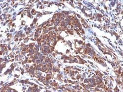
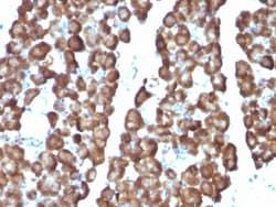
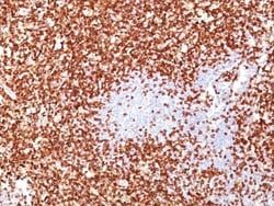
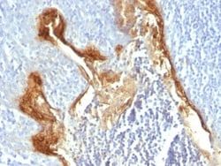
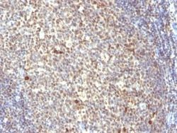
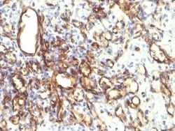
This MAb reacts with a protein of 20-30kDa, identified as PGP9.5, also known as ubiquitin carboxyl-terminal hydrolase-1 (UchL1). Initially, PGP9.5 expression in normal tissues was reported in neurons and neuroendocrine cells but later it was found in distal renal tubular epithelium, spermatogonia, Leydig cells, oocytes, melanocytes, prostatic secretory epithelium, ejaculatory duct cells, epididymis, mammary epithelial cells, Merkel cells, and dermal fibroblasts. Furthermore, immunostaining for PGP9.5 has been shown in a wide variety of mesenchymal neoplasms as well. A mutation in PGP9.5 gene is believed to cause a form of Parkinson's disease.
Related Products
Description
- Ensure accurate, reproducible results in Western Blot, Immunofluorescence UCH-L1/PGP9.5 Monoclonal specifically detects UCH-L1/PGP9.5 in Human samples
- It is validated for Western Blot.
Compare Similar Items
Show Difference

Antigen: UCH-L1/PGP9.5
Concentration: 0.2 mg/mL
Applications: Western Blot, Immunofluorescence
Conjugate: Unconjugated
Host Species: Mouse
Research Discipline: Alzheimers Research, Cellular Markers, Neurodegeneration, Neuronal Cell Markers, Neuroscience, Neurotransmission
Formulation: 10mM PBS and 0.05% BSA with 0.05% Sodium Azide
Gene ID (Entrez): 7345
Immunogen: Recombinant human UCHL1 protein
Primary or Secondary: Primary
Content And Storage: Store at 4C.
Clone: UCHL1/841
Dilution: Western Blot 0.5 - 1.0 ug/ml, Immunofluorescence 1 - 2 ug/ml
Classification: Monoclonal
Form: Purified
Regulatory Status: RUO
Target Species: Human
Gene Alias: EC 3.4.19.12, EC 6.-, Neuron cytoplasmic protein 9.5, PARK5, PGP 9.5, PGP9.5, PGP9.5Uch-L1, PGP95, ubiquitin carboxyl-terminal esterase L1 (ubiquitin thiolesterase), ubiquitin carboxyl-terminal hydrolase isozyme L1, ubiquitin C-terminal hydrolase, Ubiquitin thioesterase L1, UCHL1, UCH-L1
Gene Symbols: UCHL1
Isotype: IgG2a κ
Purification Method: Protein A or G purified
Test Specificity: This MAb reacts with a protein of 20-30kDa, identified as PGP9.5, also known as ubiquitin carboxyl-terminal hydrolase-1 (UchL1). Initially, PGP9.5 expression in normal tissues was reported in neurons and neuroendocrine cells but later it was found in distal renal tubular epithelium, spermatogonia, Leydig cells, oocytes, melanocytes, prostatic secretory epithelium, ejaculatory duct cells, epididymis, mammary epithelial cells, Merkel cells, and dermal fibroblasts. Furthermore, immunostaining for PGP9.5 has been shown in a wide variety of mesenchymal neoplasms as well. A mutation in PGP9.5 gene is believed to cause a form of Parkinson's disease.

Antigen: Phospho-Tyrosine
Concentration: 0.2 mg/mL
Applications: Flow Cytometry, Immunohistochemistry (Paraffin), SDS-Page, Immunofluorescence
Conjugate: Unconjugated
Host Species: Mouse
Research Discipline: __
Formulation: 10mM PBS and 0.05% BSA with 0.05% Sodium Azide
Gene ID (Entrez): __
Immunogen: Phosphotyrosine conjugated to BSA
Primary or Secondary: Primary
Content And Storage: Store at 4C.
Clone: PY793
Dilution: Flow Cytometry 0.5 - 1 ug/million cells in 0.1 ml, Immunohistochemistry-Paraffin 1 - 2 ug/ml, SDS-Page, Immunofluorescence 1 - 2 ug/ml
Classification: Monoclonal
Form: Purified
Regulatory Status: RUO
Target Species: Human, All species
Gene Alias: 2 amino 3(4 hydroxyphenyl) propanoic acid, 4 hydroxyphenylalanine, phosphotyrosine, pTyrosine, Tyrosine
Gene Symbols: __
Isotype: IgG2b
Purification Method: Protein A or G purified
Test Specificity: Protein phosphorylation is a fundamental event in the regulation of a large number of intracellular processes. Phosphorylation of specific tyrosine residues is the result of activation or stimulation of their respective protein tyrosine kinases. The phosphorylated proteins can be auto-phosphorylated kinases or certain cellular protein substrates. Tyrosine-phosphorylated proteins are involved in signal transduction and in the regulation of cell proliferation. Antibody to phosphotyrosine provides an excellent tool for the detection, characterization, and purification of phosphotyrosine containing proteins. This MAb shows no cross-reaction with other phosphoamino acids and is superb for multiple applications including staining of formalin/paraffin tissues.
Antigen: Podocalyxin Like
Concentration: 0.2mg/mL
Applications: Flow Cytometry, Immunohistochemistry (Paraffin), Immunofluorescence
Conjugate: Unconjugated
Host Species: Mouse
Research Discipline: Cancer, Cell Biology, Cellular Markers, Embryonic Stem Cell Markers, Hematopoietic Stem Cell Markers, Stem Cell Markers
Formulation: 10mM PBS and 0.05% BSA with 0.05% Sodium Azide
Gene ID (Entrez): 5420
Immunogen: A recombinant protein fragment containing the intracellular, transmembrane, and part of the extracellular domain of human podocalyxin.
Primary or Secondary: Primary
Content And Storage: Store at 4C.
Clone: 2A4
Dilution: Flow Cytometry 5 - 10 ul/million cells in 0.1ml, Immunohistochemistry-Paraffin 1:50 - 1:100, Immunofluorescence 1:25 - 1:50
Classification: Monoclonal
Form: Purified
Regulatory Status: RUO
Target Species: Human, Rat, Rabbit
Gene Alias: Gp200MGC138240, PCLP1, PCLP-1, PCLPGCTM-2 antigen, PCpodocalyxin, podocalyxin-like, Podocalyxin-like protein 1
Gene Symbols: PODXL
Isotype: IgM
Purification Method: Protein A or G purified
Test Specificity: Podocalyxin is a member of the CD34 transmembrane sialomucin family. It is over-expressed on the podocyte foot projections and plays essential roles in kidney development and homeostasis, blood filtration and urine formation. It is also expressed on vascular endothelia, hematopoietic progenitors and a subset of neurons. Overexpression of podocalyxin may be linked to more aggressive tumor behavior. Podocalyxin antibody can identify podocytes in the urine (podocyturia) that may indicate glomerular disease, pre-eclampsia, and other kidney pathology.