MilliporeSigma™ anti-N3-Phosphohistidine (3-pHis) Clone: SC56-2, Rabbit Monoclonal,
Manufacturer: MilliporeSigma™
Select a Size
| Pack Size | SKU | Availability | Price |
|---|---|---|---|
| Each of 1 | MABS1352MI-Each-of-1 | In Stock | ₹ 48,594.00 |
MABS1352MI - Each of 1
In Stock
Quantity
1
Base Price: ₹ 48,594.00
GST (18%): ₹ 8,746.92
Total Price: ₹ 57,340.92
Antigen
N3-Phosphohistidine (3-pHis)
Classification
Monoclonal
Conjugate
Unconjugated
Host Species
Rabbit
Purification Method
Protein A purified
Regulatory Status
RUO
Primary or Secondary
Primary
Target Species
E. coli, Human
Form
Purified
Applications
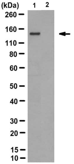
Dot Blot, Western Blot
Clone
SC56-2
Formulation
Purified rabbit monoclonal antibody in buffer containing 0.1M Tris-Glycine (pH 7.4), 150mM NaCl with 0.05% sodium azide.
Immunogen
KLH-conjugated library of random peptides containing non-hydrolyzable phosphohistidine analogue 3-pTza.
Quantity
100 μL
Research Discipline
Signaling
Test Specificity
Selectively detects proteins with histidine(s) phosphorylated at N3 of the imidazole ring (3-pHis), but not 1-pHis. Histidine phosphorylation is heat and acid labile. To generate negative control for specificity test, an aliquot of sample can be heated at 95°C for 10 to 15 minutes to reverse histidine phosphorylation. Alternatively, an aliquot of sample can be incubated under acidified pH at 37°C for 15 minutes to reduce histidine phosphorylation. Acidify each 100μL sample with 25μL of 1M HCl before the incubation, then neutralize with 25μL of 1M NaOH prior to phosphohistidine detection.
Content And Storage
2°C to 8°C for one year from date of shipment
Isotype
IgG
Related Products


Description
- Specifically detects N3-Phosphohistidine (3-pHis) Clone: SC56-2 in E
- coli, Human samples, and it is validated for Dot Blot, Western Blotting Phosphorylation plays an important role in regulating protein activities and various cellular signaling events in cells
- Limited by the tools available for phosphohistidine (pHis) detection, the majority of studies focus on serine, threonine, and tyrosine phosphorylations
- Histidine phosphorylation can occur at either N1 (1-pHis) or N3 (3-pHis) of the imidazole ring
- The development of peptides containing stable phosphoryltriazolylalanine analogues of 1-pHis and 3-pHis (1-pTza and 3-pTza) allows the generation of antibodies for studying both histidine N1 and N3 phosphorylations in signaling events
- There is growing evidence implicating His kinases in cancer and tumor metastasis and the first metastasis suppressor gene identified is one of the two known mammalian His kinases, Nm23-H1 (also known as NME1, nucleoside diphosphate kinase, or NDPK-A)
- Nm23-H1/NME1 and the closely related Nm23-H2 (NME2/NDPK-B) catalyze the transfer of phosphate from ATP onto Nucleoside-diphosphates (NDPs) through a 1-pHis enzyme intermediate
- Nm23-H1/-H2 also possess His kinase activity, transferring the phosphate from the active site pHis onto a His in a target protein
- Metabolic enzymes such as phosphoglycerate mutase (PGAM), succinyl CoA synthase (SCS), and ATP citrate lyase (ACL) also use pHis as an enzyme intermediate
- Unlike NME1/2, PGAM uses 3-pHis as an enzyme intermediate
- In addition to eukaryotes, histidine phosphorylation is well documented in bacterial 'two-component' signaling pathways involved in chemotaxis, although the phosphate is transferred from the pHis formed in the receptor/sensor protein to Asp residues of an acceptor response regulator protein, and the receptor/sensor protein essentially functions as an aspartate kinase.
Compare Similar Items
Show Difference

Antigen: N3-Phosphohistidine (3-pHis)
Classification: Monoclonal
Conjugate: Unconjugated
Host Species: Rabbit
Purification Method: Protein A purified
Regulatory Status: RUO
Primary or Secondary: Primary
Target Species: E. coli, Human
Form: Purified
Applications: Dot Blot, Western Blot
Clone: SC56-2
Formulation: Purified rabbit monoclonal antibody in buffer containing 0.1M Tris-Glycine (pH 7.4), 150mM NaCl with 0.05% sodium azide.
Immunogen: KLH-conjugated library of random peptides containing non-hydrolyzable phosphohistidine analogue 3-pTza.
Quantity: 100 μL
Research Discipline: Signaling
Test Specificity: Selectively detects proteins with histidine(s) phosphorylated at N3 of the imidazole ring (3-pHis), but not 1-pHis. Histidine phosphorylation is heat and acid labile. To generate negative control for specificity test, an aliquot of sample can be heated at 95°C for 10 to 15 minutes to reverse histidine phosphorylation. Alternatively, an aliquot of sample can be incubated under acidified pH at 37°C for 15 minutes to reduce histidine phosphorylation. Acidify each 100μL sample with 25μL of 1M HCl before the incubation, then neutralize with 25μL of 1M NaOH prior to phosphohistidine detection.
Content And Storage: 2°C to 8°C for one year from date of shipment
Isotype: IgG

Antigen: RasGRP1
Classification: Monoclonal
Conjugate: Unconjugated
Host Species: Mouse
Purification Method: Protein G purified
Regulatory Status: __
Primary or Secondary: Primary
Target Species: Human
Form: Purified
Applications: Western Blot
Clone: 10.1
Formulation: Purified mouse monoclonal IgG2aκ in buffer containing 0.1MTris-Glycine (pH 7.4), 150mM NaCl with 0.05% sodium azide.
Immunogen: GST-tagged recombinant protein corresponding to human RasGRP1.
Quantity: 100 μg
Research Discipline: __
Test Specificity: __
Content And Storage: 2-8°C for one year
Isotype: IgG2a κ

Antigen: SGK1
Classification: Monoclonal
Conjugate: Unconjugated
Host Species: Mouse
Purification Method: Protein G Purified
Regulatory Status: RUO
Primary or Secondary: Primary
Target Species: Human
Form: Purified
Applications: Western Blot
Clone: 1G2.1
Formulation: Purified mouse IgG1 in buffer containing 0.1M Tris-Glycine (pH 7.4), 150mM NaCl with 0.05% Sodium Azide.
Immunogen: GST-tagged recombinant human SGK1 fragment corresponding to the region N-terminal to the kinase domain.
Quantity: 100 μg
Research Discipline: Signaling
Test Specificity: Clone 1G2.1 was raised against SGK1 N-terminal fragment, reactivity towards human spliced isoforms 2-5 and murine spliced isoforms 2 and 3 are possible only if the epitope lies outside the first 25-amino acids of isoform 1.
Content And Storage: Stable for 1 year at 2°-8°C from date of receipt.
Isotype: IgG1 κ